 Movie
Movie Controller

[English] 日本語
 Yorodumi
- PDB-4ij3: Oxidoreductase Fragment of Human QSOX1 in Complex with a FAB Frag... -
+ Open data

- Basic information
| Entry | Database: PDB / ID: 4ij3 | |||||||||
|---|---|---|---|---|---|---|---|---|---|---|
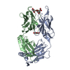
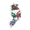
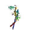
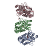
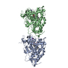
| Title | Oxidoreductase Fragment of Human QSOX1 in Complex with a FAB Fragment from an Anti- Human QSOX1 Antibody | |||||||||
 Components Components |
| |||||||||
 Keywords Keywords | OXIDOREDUCTASE/OXIDOREDUCTASE INHIBITOR / inhibitor / antibody binding / OXIDOREDUCTASE-OXIDOREDUCTASE INHIBITOR complex | |||||||||
| Function / homology |  Function and homology information Function and homology informationflavin-dependent sulfhydryl oxidase activity / thiol oxidase / extracellular matrix assembly / negative regulation of macroautophagy / protein disulfide isomerase activity / FAD binding / platelet alpha granule lumen / Post-translational protein phosphorylation / specific granule lumen / Regulation of Insulin-like Growth Factor (IGF) transport and uptake by Insulin-like Growth Factor Binding Proteins (IGFBPs) ...flavin-dependent sulfhydryl oxidase activity / thiol oxidase / extracellular matrix assembly / negative regulation of macroautophagy / protein disulfide isomerase activity / FAD binding / platelet alpha granule lumen / Post-translational protein phosphorylation / specific granule lumen / Regulation of Insulin-like Growth Factor (IGF) transport and uptake by Insulin-like Growth Factor Binding Proteins (IGFBPs) / tertiary granule lumen / Platelet degranulation / protein folding / endoplasmic reticulum lumen / Golgi membrane / Neutrophil degranulation / Golgi apparatus / : / extracellular exosome / extracellular region Similarity search - Function | |||||||||
| Biological species |  Homo sapiens (human) Homo sapiens (human) | |||||||||
| Method |  X-RAY DIFFRACTION / MOLECULAR REPLACEMENT / Resolution: 2.7 Å X-RAY DIFFRACTION / MOLECULAR REPLACEMENT / Resolution: 2.7 Å | |||||||||
 Authors Authors | Fass, D. / Grossman, I. | |||||||||
 Citation Citation | Journal: J.Mol.Biol. / Year: 2013 Title: An Inhibitory Antibody Blocks the First Step in the Dithiol/Disulfide Relay Mechanism of the Enzyme QSOX1. Authors: Grossman, I. / Alon, A. / Ilani, T. / Fass, D. | |||||||||
| History |
|
- Structure visualization
| Structure viewer | Molecule: MolmilJmol/JSmol |
|---|
- Downloads & links
-Download
| PDBx/mmCIF format | 4ij3.cif.gz | 150.4 KB | Display | PDBx/mmCIF format |
|---|---|---|---|---|
| PDB format | pdb4ij3.ent.gz | 115.8 KB | Display | PDB format |
| PDBx/mmJSON format | 4ij3.json.gz | Tree view | PDBx/mmJSON format | |
| Others |  Other downloads Other downloads |
-Validation report
| Arichive directory | https://data.pdbj.org/pub/pdb/validation_reports/ij/4ij3ftp://data.pdbj.org/pub/pdb/validation_reports/ij/4ij3 | HTTPS FTP |
|---|
-Related structure data


-Links
 PDBj
PDBj


- Assembly
| Deposited unit | 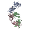
| ||||||||
|---|---|---|---|---|---|---|---|---|---|
| 1 |
| ||||||||
| Unit cell |
|
-Components
| #1: Protein | Mass: 26986.654 Da / Num. of mol.: 1 / Fragment: Oxidoreductase fragment, UNP RESIDUES 33-272 Source method: isolated from a genetically manipulated source Source: (gene. exp.) Homo sapiens (human) / Gene: QSOX1, QSCN6, UNQ2520/PRO6013 / Plasmid: PET15b / Production host:  | ||||
|---|---|---|---|---|---|
| #2: Antibody | Mass: 23639.115 Da / Num. of mol.: 1 Source method: isolated from a genetically manipulated source Source: (gene. exp.) | ||||
| #3: Antibody | Mass: 23492.381 Da / Num. of mol.: 1 Source method: isolated from a genetically manipulated source Source: (gene. exp.) | ||||
| #4: Chemical |   Mass: 94.971 Da / Num. of mol.: 2 / Source method: obtained synthetically / Formula: PO4 Mass: 94.971 Da / Num. of mol.: 2 / Source method: obtained synthetically / Formula: PO4#5: Water | ChemComp-HOH / |  Mass: 18.015 Da / Num. of mol.: 350 / Source method: isolated from a natural source / Formula: H2O Mass: 18.015 Da / Num. of mol.: 350 / Source method: isolated from a natural source / Formula: H2OHas protein modification | Y | |
-Experimental details
-Experiment
| Experiment | Method: X-RAY DIFFRACTION / Number of used crystals: 1 |
|---|
- Sample preparation
| Crystal | Density Matthews: 3.37 Å3/Da / Density % sol: 63.47 % |
|---|---|
| Crystal grow | Temperature: 293 K / Method: vapor diffusion, hanging drop / pH: 8 Details: 19% w/v PEG 4000, 0.4 M ammonium phosphate dibasic, pH 8, VAPOR DIFFUSION, HANGING DROP, temperature 293K |
-Data collection
| Diffraction | Mean temperature: 100 K |
|---|---|
| Diffraction source | Source: ROTATING ANODE / Type: RIGAKU RUH3R / Wavelength: 1.5418 Å |
| Detector | Type: RIGAKU RAXIS IV++ / Detector: IMAGE PLATE / Date: Feb 27, 2012 / Details: osmic mirrors |
| Radiation | Protocol: SINGLE WAVELENGTH / Monochromatic (M) / Laue (L): M / Scattering type: x-ray |
| Radiation wavelength | Wavelength: 1.5418 Å / Relative weight: 1 |
| Reflection | Resolution: 2.7→50 Å / Num. all: 38606 / Num. obs: 37560 / % possible obs: 97.3 % / Observed criterion σ(F): 0 / Observed criterion σ(I): -3 / Redundancy: 5.1 % / Biso Wilson estimate: 55.29 Å2 / Rmerge(I) obs: 0.092 / Rsym value: 0.092 / Net I/σ(I): 14.35 |
| Reflection shell | Resolution: 2.7→2.75 Å / Redundancy: 3.8 % / Rmerge(I) obs: 0.363 / Mean I/σ(I) obs: 1.96 / Num. unique all: 1769 |
- Processing
| Software |
| |||||||||||||||||||||||||
|---|---|---|---|---|---|---|---|---|---|---|---|---|---|---|---|---|---|---|---|---|---|---|---|---|---|---|
| Refinement | Method to determine structure: MOLECULAR REPLACEMENT Starting model: 3Q6O, 3OKD Resolution: 2.7→50 Å / Isotropic thermal model: isotropic / Cross valid method: THROUGHOUT / σ(F): 0 / Stereochemistry target values: Engh & Huber
| |||||||||||||||||||||||||
| Refinement step | Cycle: LAST / Resolution: 2.7→50 Å
| |||||||||||||||||||||||||
| Refine LS restraints |
| |||||||||||||||||||||||||
| LS refinement shell | Resolution: 2.7→2.72 Å
|
