 Movie
Movie Controller

[English] 日本語
 Yorodumi
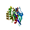
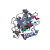
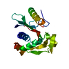
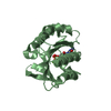
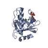
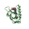
- PDB-4i03: Human MMP12 in complex with a PEG-linked bifunctional L-glutamate... -
+ Open data

- Basic information
| Entry | Database: PDB / ID: 4i03 | ||||||
|---|---|---|---|---|---|---|---|
| Title | Human MMP12 in complex with a PEG-linked bifunctional L-glutamate motif inhibitor | ||||||
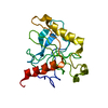
 Components Components | Macrophage metalloelastase | ||||||
 Keywords Keywords | HYDROLASE/HYDROLASE INHIBITOR / selective carboxylate based MMP-12 bifunctional inhibitor / METZINCIN / Zinc protease / HYDROLASE-HYDROLASE INHIBITOR complex | ||||||
| Function / homology |  Function and homology information Function and homology informationmacrophage elastase / negative regulation of endothelial cell-matrix adhesion / bronchiole development / positive regulation of epithelial cell proliferation involved in wound healing / elastin catabolic process / regulation of defense response to virus by host / positive regulation of type I interferon-mediated signaling pathway / wound healing, spreading of epidermal cells / negative regulation of type I interferon-mediated signaling pathway / lung alveolus development ...macrophage elastase / negative regulation of endothelial cell-matrix adhesion / bronchiole development / positive regulation of epithelial cell proliferation involved in wound healing / elastin catabolic process / regulation of defense response to virus by host / positive regulation of type I interferon-mediated signaling pathway / wound healing, spreading of epidermal cells / negative regulation of type I interferon-mediated signaling pathway / lung alveolus development / response to amyloid-beta / Collagen degradation / collagen catabolic process / positive regulation of interferon-alpha production / extracellular matrix disassembly / core promoter sequence-specific DNA binding / collagen binding / Degradation of the extracellular matrix / extracellular matrix organization / extracellular matrix / metalloendopeptidase activity / cellular response to virus / protein import into nucleus / endopeptidase activity / sequence-specific DNA binding / serine-type endopeptidase activity / calcium ion binding / negative regulation of transcription by RNA polymerase II / positive regulation of transcription by RNA polymerase II / proteolysis / extracellular space / extracellular region / zinc ion binding / nucleus / cytoplasm Similarity search - Function | ||||||
| Biological species |  Homo sapiens (human) Homo sapiens (human) | ||||||
| Method |  X-RAY DIFFRACTION / SYNCHROTRON / MOLECULAR REPLACEMENT / Resolution: 1.7 Å X-RAY DIFFRACTION / SYNCHROTRON / MOLECULAR REPLACEMENT / Resolution: 1.7 Å | ||||||
 Authors Authors | Stura, E.A. / Vera, L. / Devel, L. / Cassar-Lajeunesse, E. / Dive, V. | ||||||
 Citation Citation | Journal: J.Struct.Biol. / Year: 2013 Title: Crystallization of bi-functional ligand protein complexes. Authors: Antoni, C. / Vera, L. / Devel, L. / Catalani, M.P. / Czarny, B. / Cassar-Lajeunesse, E. / Nuti, E. / Rossello, A. / Dive, V. / Stura, E.A. #1: Journal: Bioorg.Med.Chem.Lett. / Year: 2005 Title: A new development of matrix metalloproteinase inhibitors: twin hydroxamic acids as potent inhibitors of MMPs. Authors: Rossello, A. / Nuti, E. / Catalani, M.P. / Carelli, P. / Orlandini, E. / Rapposelli, S. / Tuccinardi, T. / Atkinson, S.J. / Murphy, G. / Balsamo, A. | ||||||
| History |
|
- Structure visualization
| Structure viewer | Molecule: MolmilJmol/JSmol |
|---|
- Downloads & links
-Download
| PDBx/mmCIF format | 4i03.cif.gz | 59.4 KB | Display | PDBx/mmCIF format |
|---|---|---|---|---|
| PDB format | pdb4i03.ent.gz | 41.2 KB | Display | PDB format |
| PDBx/mmJSON format | 4i03.json.gz | Tree view | PDBx/mmJSON format | |
| Others |  Other downloads Other downloads |
-Validation report
| Arichive directory | https://data.pdbj.org/pub/pdb/validation_reports/i0/4i03ftp://data.pdbj.org/pub/pdb/validation_reports/i0/4i03 | HTTPS FTP |
|---|
-Related structure data
| Related structure data | 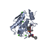 4h1qC 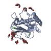 4h2eC  4h30C 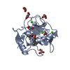 4h3xC  4h49C 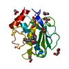 4h76C 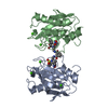 4h82C  4h84C 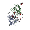 4hmaC C: citing same article ( |
|---|---|
| Similar structure data |


-Links
 PDBj
PDBj


- Assembly
| Deposited unit | 
| ||||||||
|---|---|---|---|---|---|---|---|---|---|
| 1 |
| ||||||||
| Unit cell |
| ||||||||
| Components on special symmetry positions |
| ||||||||
| Details | The bifunctional inhibitor joins two MMP-12 molecules creating a crystallographic dimer |
-Components
-Protein , 1 types, 1 molecules A
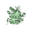
| #1: Protein | Mass: 17557.633 Da / Num. of mol.: 1 / Fragment: catalytic domain (UNP residues 106-263) / Mutation: F171D, E219A Source method: isolated from a genetically manipulated source Source: (gene. exp.) Homo sapiens (human) / Strain: Escherichia coli / Gene: HME, MMP12 / Plasmid: PET24A / Production host:  |
|---|
-Non-polymers , 9 types, 189 molecules 


| #2: Chemical |  Mass: 65.409 Da / Num. of mol.: 2 / Source method: obtained synthetically / Formula: Zn Mass: 65.409 Da / Num. of mol.: 2 / Source method: obtained synthetically / Formula: Zn#3: Chemical |  Mass: 40.078 Da / Num. of mol.: 3 / Source method: obtained synthetically / Formula: Ca Mass: 40.078 Da / Num. of mol.: 3 / Source method: obtained synthetically / Formula: Ca#4: Chemical | ChemComp-L88 / ( |  Mass: 1059.295 Da / Num. of mol.: 1 / Source method: obtained synthetically / Formula: C58H66N4O11S2 Mass: 1059.295 Da / Num. of mol.: 1 / Source method: obtained synthetically / Formula: C58H66N4O11S2#5: Chemical |  Mass: 92.094 Da / Num. of mol.: 3 / Source method: obtained synthetically / Formula: C3H8O3 Mass: 92.094 Da / Num. of mol.: 3 / Source method: obtained synthetically / Formula: C3H8O3#6: Chemical |  Mass: 76.094 Da / Num. of mol.: 2 / Source method: obtained synthetically / Formula: C3H8O2 Mass: 76.094 Da / Num. of mol.: 2 / Source method: obtained synthetically / Formula: C3H8O2#7: Chemical |  Mass: 78.133 Da / Num. of mol.: 2 / Source method: obtained synthetically / Formula: C2H6OS / Comment: DMSO, precipitant*YM Mass: 78.133 Da / Num. of mol.: 2 / Source method: obtained synthetically / Formula: C2H6OS / Comment: DMSO, precipitant*YM#8: Chemical | ChemComp-EDO /  Mass: 62.068 Da / Num. of mol.: 4 / Source method: obtained synthetically / Formula: C2H6O2 Mass: 62.068 Da / Num. of mol.: 4 / Source method: obtained synthetically / Formula: C2H6O2#9: Chemical | ChemComp-PEG / |  Mass: 106.120 Da / Num. of mol.: 1 / Source method: obtained synthetically / Formula: C4H10O3 Mass: 106.120 Da / Num. of mol.: 1 / Source method: obtained synthetically / Formula: C4H10O3#10: Water | ChemComp-HOH / | Mass: 18.015 Da / Num. of mol.: 171 / Source method: isolated from a natural source / Formula: H2O |
|---|
-Experimental details
-Experiment
| Experiment | Method: X-RAY DIFFRACTION / Number of used crystals: 1 |
|---|
- Sample preparation
| Crystal | Density Matthews: 2.18 Å3/Da / Density % sol: 43.45 % |
|---|---|
| Crystal grow | Temperature: 293 K / Method: vapor diffusion, sitting drop / pH: 8.5 Details: Protein solution: 222 microM MMP12 mutant E219A 106 microM bifunctional inhibitor LD884. Reservoir: 27% PEG 10K, 150mM imidazole piperidine, pH 8.5. Cryoprotectant: 5 % di-ethylene glycol + ...Details: Protein solution: 222 microM MMP12 mutant E219A 106 microM bifunctional inhibitor LD884. Reservoir: 27% PEG 10K, 150mM imidazole piperidine, pH 8.5. Cryoprotectant: 5 % di-ethylene glycol + 5 % ethylene glycol + 10 % 1,2-propanediol + 5 % DMSO + 5 % glycerol, 25% MPEG 5K, 100mM (Na acetate, ADA, Bicine 10% pH 4.0/90% pH 9.0), VAPOR DIFFUSION, SITTING DROP, temperature 293K |
-Data collection
| Diffraction | Mean temperature: 100 K | ||||||||||||||||||||||||||||||||||||||||||||||||||||||||||||||||||||||
|---|---|---|---|---|---|---|---|---|---|---|---|---|---|---|---|---|---|---|---|---|---|---|---|---|---|---|---|---|---|---|---|---|---|---|---|---|---|---|---|---|---|---|---|---|---|---|---|---|---|---|---|---|---|---|---|---|---|---|---|---|---|---|---|---|---|---|---|---|---|---|---|
| Diffraction source | Source: SYNCHROTRON / Site: SOLEIL  / Beamline: PROXIMA 1 / Wavelength: 0.98011 Å / Beamline: PROXIMA 1 / Wavelength: 0.98011 Å | ||||||||||||||||||||||||||||||||||||||||||||||||||||||||||||||||||||||
| Detector | Type: PSI PILATUS 6M / Detector: PIXEL / Date: Oct 12, 2012 / Details: mirrors | ||||||||||||||||||||||||||||||||||||||||||||||||||||||||||||||||||||||
| Radiation | Monochromator: SAGITALLY FOCUSED Si(111) / Protocol: SINGLE WAVELENGTH / Monochromatic (M) / Laue (L): M / Scattering type: x-ray | ||||||||||||||||||||||||||||||||||||||||||||||||||||||||||||||||||||||
| Radiation wavelength | Wavelength: 0.98011 Å / Relative weight: 1 | ||||||||||||||||||||||||||||||||||||||||||||||||||||||||||||||||||||||
| Reflection | Resolution: 1.7→50 Å / Num. all: 16789 / Num. obs: 16663 / % possible obs: 99.2 % / Observed criterion σ(F): 0 / Observed criterion σ(I): -4 / Redundancy: 4.14 % / Biso Wilson estimate: 28.35 Å2 / Rmerge(I) obs: 0.102 / Rsym value: 0.089 / Net I/σ(I): 9.52 | ||||||||||||||||||||||||||||||||||||||||||||||||||||||||||||||||||||||
| Reflection shell | Diffraction-ID: 1
|
- Processing
| Software |
| |||||||||||||||||||||||||||||||||||||||||||||||||||||||||||||||||||||||||||||||||||||
|---|---|---|---|---|---|---|---|---|---|---|---|---|---|---|---|---|---|---|---|---|---|---|---|---|---|---|---|---|---|---|---|---|---|---|---|---|---|---|---|---|---|---|---|---|---|---|---|---|---|---|---|---|---|---|---|---|---|---|---|---|---|---|---|---|---|---|---|---|---|---|---|---|---|---|---|---|---|---|---|---|---|---|---|---|---|---|
| Refinement | Method to determine structure: MOLECULAR REPLACEMENT / Resolution: 1.7→36.67 Å / Cor.coef. Fo:Fc: 0.974 / Cor.coef. Fo:Fc free: 0.96 / SU B: 2.294 / SU ML: 0.074 / Isotropic thermal model: Isotropic / Cross valid method: THROUGHOUT / σ(F): 0 / σ(I): -4 / ESU R: 0.104 / ESU R Free: 0.108 / Stereochemistry target values: MAXIMUM LIKELIHOOD / Details: HYDROGENS HAVE BEEN ADDED IN THE RIDING POSITIONS
| |||||||||||||||||||||||||||||||||||||||||||||||||||||||||||||||||||||||||||||||||||||
| Solvent computation | Ion probe radii: 0.8 Å / Shrinkage radii: 0.8 Å / VDW probe radii: 1.4 Å / Solvent model: MASK | |||||||||||||||||||||||||||||||||||||||||||||||||||||||||||||||||||||||||||||||||||||
| Displacement parameters | Biso mean: 19.56 Å2
| |||||||||||||||||||||||||||||||||||||||||||||||||||||||||||||||||||||||||||||||||||||
| Refinement step | Cycle: LAST / Resolution: 1.7→36.67 Å
| |||||||||||||||||||||||||||||||||||||||||||||||||||||||||||||||||||||||||||||||||||||
| Refine LS restraints |
| |||||||||||||||||||||||||||||||||||||||||||||||||||||||||||||||||||||||||||||||||||||
| LS refinement shell | Resolution: 1.696→1.74 Å / Total num. of bins used: 20
|
