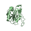
response to brefeldin A / regulation of auxin metabolic process / vesicle-mediated transport to the plasma membrane / auxin-activated signaling pathway / phospholipase activator activity / vesicle-mediated transport / small monomeric GTPase / trans-Golgi network / intracellular protein transport / G protein activity ...response to brefeldin A / regulation of auxin metabolic process / vesicle-mediated transport to the plasma membrane / auxin-activated signaling pathway / phospholipase activator activity / vesicle-mediated transport / small monomeric GTPase / trans-Golgi network / intracellular protein transport / G protein activity / early endosome / mRNA binding / GTP binding / Golgi apparatus / cytosol / cytoplasm Similarity search - Function
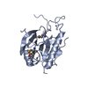
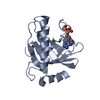
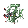
ADP-ribosylation factor 1-5 / Small GTPase superfamily, ARF type / Small GTPase Arf domain profile. / Sar1p-like members of the Ras-family of small GTPases / Small GTPase superfamily, ARF/SAR type / ADP-ribosylation factor family / ARF-like small GTPases; ARF, ADP-ribosylation factor / Rab subfamily of small GTPases / Small GTP-binding protein domain / P-loop containing nucleotide triphosphate hydrolases ...ADP-ribosylation factor 1-5 / Small GTPase superfamily, ARF type / Small GTPase Arf domain profile. / Sar1p-like members of the Ras-family of small GTPases / Small GTPase superfamily, ARF/SAR type / ADP-ribosylation factor family / ARF-like small GTPases; ARF, ADP-ribosylation factor / Rab subfamily of small GTPases / Small GTP-binding protein domain / P-loop containing nucleotide triphosphate hydrolases / Rossmann fold / P-loop containing nucleoside triphosphate hydrolase / 3-Layer(aba) Sandwich / Alpha Beta Similarity search - Domain/homology
Method to determine structure: MOLECULAR REPLACEMENT / Resolution: 1.8→89.8 Å / Cor.coef. Fo:Fc: 0.947 / Cor.coef. Fo:Fc free: 0.917 / SU B: 4.81 / SU ML: 0.086 / Cross valid method: THROUGHOUT / ESU R Free: 0.134 / Stereochemistry target values: MAXIMUM LIKELIHOOD / Details: HYDROGENS HAVE BEEN ADDED IN THE RIDING POSITIONS
Rfactor
Num. reflection
% reflection
Selection details
Rfree
0.24148
1750
5 %
RANDOM
Rwork
0.19858
-
-
-
obs
0.20076
33083
99.69 %
-
Solvent computation
Ion probe radii: 0.8 Å / Shrinkage radii: 0.8 Å / VDW probe radii: 1.2 Å / Solvent model: MASK
Displacement parameters
Biso mean: 26.032 Å2
Baniso -1
Baniso -2
Baniso -3
1-
-0.79 Å2
0 Å2
0 Å2
2-
-
-0.21 Å2
0 Å2
3-
-
-
1.01 Å2
Refinement step
Cycle: LAST / Resolution: 1.8→89.8 Å
Protein
Nucleic acid
Ligand
Solvent
Total
Num. atoms
2665
0
58
290
3013
Refine LS restraints
Refine-ID
Type
Dev ideal
Dev ideal target
Number
X-RAY DIFFRACTION
r_bond_refined_d
0.015
0.022
2770
X-RAY DIFFRACTION
r_angle_refined_deg
1.496
1.976
3753
X-RAY DIFFRACTION
r_dihedral_angle_1_deg
6.081
5
329
X-RAY DIFFRACTION
r_dihedral_angle_2_deg
32.567
24.154
130
X-RAY DIFFRACTION
r_dihedral_angle_3_deg
14.529
15
491
X-RAY DIFFRACTION
r_dihedral_angle_4_deg
19.401
15
19
X-RAY DIFFRACTION
r_chiral_restr
0.099
0.2
426
X-RAY DIFFRACTION
r_gen_planes_refined
0.006
0.02
2032
X-RAY DIFFRACTION
r_nbd_refined
0.209
0.2
1208
X-RAY DIFFRACTION
r_nbtor_refined
0.309
0.2
1855
X-RAY DIFFRACTION
r_xyhbond_nbd_refined
0.139
0.2
217
X-RAY DIFFRACTION
r_metal_ion_refined
0.032
0.2
1
X-RAY DIFFRACTION
r_symmetry_vdw_refined
0.342
0.2
61
X-RAY DIFFRACTION
r_symmetry_hbond_refined
0.175
0.2
16
X-RAY DIFFRACTION
r_mcbond_it
1.028
1.5
1692
X-RAY DIFFRACTION
r_mcangle_it
1.616
2
2642
X-RAY DIFFRACTION
r_scbond_it
2.482
3
1255
X-RAY DIFFRACTION
r_scangle_it
3.852
4.5
1111
LS refinement shell
Resolution: 1.8→1.847 Å / Total num. of bins used: 20
Rfactor
Num. reflection
% reflection
Rfree
0.284
129
-
Rwork
0.218
2413
-
obs
-
-
99.57 %
Refinement TLS params.
Method: refined / Origin x: 29.1465 Å / Origin y: 12.3102 Å / Origin z: 11.3442 Å
11
12
13
21
22
23
31
32
33
T
-0.1132 Å2
0.0059 Å2
0.014 Å2
-
-0.107 Å2
-0.034 Å2
-
-
-0.1083 Å2
L
1.6879 °2
1.4822 °2
-0.6108 °2
-
1.8466 °2
-0.6418 °2
-
-
0.4141 °2
S
-0.1182 Å °
0.1039 Å °
-0.1576 Å °
-0.161 Å °
0.0718 Å °
-0.1166 Å °
0.0408 Å °
0.0139 Å °
0.0464 Å °
Refinement TLS group
ID
Refine-ID
Refine TLS-ID
Auth asym-ID
Auth seq-ID
1
X-RAY DIFFRACTION
1
A
4 - 181
2
X-RAY DIFFRACTION
1
B
4 - 181
+
About Yorodumi
-
News
-
Feb 9, 2022. New format data for meta-information of EMDB entries
New format data for meta-information of EMDB entries
Version 3 of the EMDB header file is now the official format.
The previous official version 1.9 will be removed from the archive.
In the structure databanks used in Yorodumi, some data are registered as the other names, "COVID-19 virus" and "2019-nCoV". Here are the details of the virus and the list of structure data.
Jan 31, 2019. EMDB accession codes are about to change! (news from PDBe EMDB page)
EMDB accession codes are about to change! (news from PDBe EMDB page)
The allocation of 4 digits for EMDB accession codes will soon come to an end. Whilst these codes will remain in use, new EMDB accession codes will include an additional digit and will expand incrementally as the available range of codes is exhausted. The current 4-digit format prefixed with “EMD-” (i.e. EMD-XXXX) will advance to a 5-digit format (i.e. EMD-XXXXX), and so on. It is currently estimated that the 4-digit codes will be depleted around Spring 2019, at which point the 5-digit format will come into force.
The EM Navigator/Yorodumi systems omit the EMD- prefix.
Related info.:Q: What is EMD? / ID/Accession-code notation in Yorodumi/EM Navigator
Yorodumi is a browser for structure data from EMDB, PDB, SASBDB, etc.
This page is also the successor to EM Navigator detail page, and also detail information page/front-end page for Omokage search.
The word "yorodu" (or yorozu) is an old Japanese word meaning "ten thousand". "mi" (miru) is to see.
Related info.:EMDB / PDB / SASBDB / Comparison of 3 databanks / Yorodumi Search / Aug 31, 2016. New EM Navigator & Yorodumi / Yorodumi Papers / Jmol/JSmol / Function and homology information / Changes in new EM Navigator and Yorodumi
 Movie
Movie Controller

 Yorodumi
 Open data
 Basic information
 Components
 Keywords
 Function and homology information
 X-RAY DIFFRACTION /
  Authors
 Citation
 Structure visualization
 Downloads & links
 Other downloads
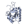


 PDBj
PDBj


 Assembly
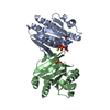

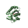


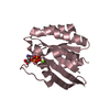
 Mass: 24.305 Da / Num. of mol.: 2 / Source method: obtained synthetically / Formula: Mg
 Type: RNA linking / Mass: 443.201 Da / Num. of mol.: 2 / Source method: obtained synthetically / Formula: C10H15N5O11P2 / Comment: GDP, energy-carrying molecule*YM
 Mass: 18.015 Da / Num. of mol.: 290 / Source method: isolated from a natural source / Formula: H2O
 Sample preparation
 / Beamline: X29A
 Processing