 Movie
Movie Controller

[English] 日本語
 Yorodumi
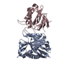
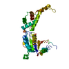
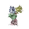
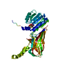
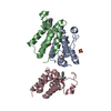
- PDB-4hrl: Structural Basis for Eliciting a Cytotoxic Effect in HER2-Overexp... -
+ Open data

- Basic information
| Entry | Database: PDB / ID: 4hrl | ||||||
|---|---|---|---|---|---|---|---|
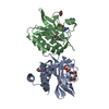
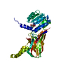
| Title | Structural Basis for Eliciting a Cytotoxic Effect in HER2-Overexpressing Cancer Cells via Binding to the Extracellular Domain of HER2 | ||||||
 Components Components |
| ||||||
 Keywords Keywords | TRANSFERASE/DE NOVO PROTEIN / TRANSFERASE-DE NOVO PROTEIN complex | ||||||
| Function / homology |  Function and homology information Function and homology informationnegative regulation of immature T cell proliferation in thymus / ERBB3:ERBB2 complex / ERBB2-ERBB4 signaling pathway / immature T cell proliferation in thymus / GRB7 events in ERBB2 signaling / RNA polymerase I core binding / semaphorin receptor complex / Developmental Lineage of Mammary Stem Cells / ErbB-3 class receptor binding / motor neuron axon guidance ...negative regulation of immature T cell proliferation in thymus / ERBB3:ERBB2 complex / ERBB2-ERBB4 signaling pathway / immature T cell proliferation in thymus / GRB7 events in ERBB2 signaling / RNA polymerase I core binding / semaphorin receptor complex / Developmental Lineage of Mammary Stem Cells / ErbB-3 class receptor binding / motor neuron axon guidance / Sema4D induced cell migration and growth-cone collapse / regulation of microtubule-based process / PLCG1 events in ERBB2 signaling / ERBB2-EGFR signaling pathway / enzyme-linked receptor protein signaling pathway / ERBB2 Activates PTK6 Signaling / ERBB2-ERBB3 signaling pathway / Drug-mediated inhibition of ERBB2 signaling / Resistance of ERBB2 KD mutants to trastuzumab / Resistance of ERBB2 KD mutants to sapitinib / Resistance of ERBB2 KD mutants to tesevatinib / Resistance of ERBB2 KD mutants to neratinib / Resistance of ERBB2 KD mutants to osimertinib / Resistance of ERBB2 KD mutants to afatinib / Resistance of ERBB2 KD mutants to AEE788 / Resistance of ERBB2 KD mutants to lapatinib / Drug resistance in ERBB2 TMD/JMD mutants / neurotransmitter receptor localization to postsynaptic specialization membrane / positive regulation of Rho protein signal transduction / positive regulation of MAP kinase activity / oligodendrocyte differentiation / positive regulation of transcription by RNA polymerase I / ERBB2 Regulates Cell Motility / Developmental Lineage of Mammary Gland Myoepithelial Cells / semaphorin-plexin signaling pathway / neuromuscular junction development / PI3K events in ERBB2 signaling / regulation of angiogenesis / positive regulation of protein targeting to membrane / regulation of ERK1 and ERK2 cascade / Schwann cell development / coreceptor activity / Signaling by ERBB2 / TFAP2 (AP-2) family regulates transcription of growth factors and their receptors / myelination / peptidyl-tyrosine phosphorylation / transmembrane receptor protein tyrosine kinase activity / positive regulation of epithelial cell proliferation / GRB2 events in ERBB2 signaling / positive regulation of cell adhesion / SHC1 events in ERBB2 signaling / cell surface receptor protein tyrosine kinase signaling pathway / cellular response to epidermal growth factor stimulus / basal plasma membrane / Constitutive Signaling by Overexpressed ERBB2 / Downregulation of ERBB2:ERBB3 signaling / positive regulation of translation / phosphatidylinositol 3-kinase/protein kinase B signal transduction / neuromuscular junction / wound healing / Signaling by ERBB2 TMD/JMD mutants / receptor protein-tyrosine kinase / Signaling by ERBB2 ECD mutants / Signaling by ERBB2 KD Mutants / cellular response to growth factor stimulus / receptor tyrosine kinase binding / epidermal growth factor receptor signaling pathway / ruffle membrane / Downregulation of ERBB2 signaling / neuron differentiation / Constitutive Signaling by Aberrant PI3K in Cancer / transmembrane signaling receptor activity / PIP3 activates AKT signaling / myelin sheath / heart development / PI5P, PP2A and IER3 Regulate PI3K/AKT Signaling / positive regulation of cell growth / RAF/MAP kinase cascade / protein tyrosine kinase activity / presynaptic membrane / basolateral plasma membrane / protein phosphorylation / positive regulation of MAPK cascade / early endosome / cell population proliferation / cell surface receptor signaling pathway / signaling receptor complex / apical plasma membrane / endosome membrane / intracellular signal transduction / protein heterodimerization activity / signaling receptor binding / negative regulation of apoptotic process / perinuclear region of cytoplasm / signal transduction / nucleoplasm / ATP binding / membrane / identical protein binding / nucleus Similarity search - Function | ||||||
| Biological species |  Homo sapiens (human) Homo sapiens (human)synthetic (others) | ||||||
| Method |  X-RAY DIFFRACTION / SYNCHROTRON / MOLECULAR REPLACEMENT / molecular replacement / Resolution: 2.55 Å X-RAY DIFFRACTION / SYNCHROTRON / MOLECULAR REPLACEMENT / molecular replacement / Resolution: 2.55 Å | ||||||
 Authors Authors | Jost, C. / Schilling, J. / Plueckthun, A. | ||||||
 Citation Citation | Journal: Structure / Year: 2013 Title: Structural Basis for Eliciting a Cytotoxic Effect in HER2-Overexpressing Cancer Cells via Binding to the Extracellular Domain of HER2. Authors: Jost, C. / Schilling, J. / Tamaskovic, R. / Schwill, M. / Honegger, A. / Plueckthun, A. | ||||||
| History |
|
- Structure visualization
| Structure viewer | Molecule: MolmilJmol/JSmol |
|---|
- Downloads & links
-Download
| PDBx/mmCIF format | 4hrl.cif.gz | 77 KB | Display | PDBx/mmCIF format |
|---|---|---|---|---|
| PDB format | pdb4hrl.ent.gz | 56 KB | Display | PDB format |
| PDBx/mmJSON format | 4hrl.json.gz | Tree view | PDBx/mmJSON format | |
| Others |  Other downloads Other downloads |
-Validation report
| Arichive directory | https://data.pdbj.org/pub/pdb/validation_reports/hr/4hrlftp://data.pdbj.org/pub/pdb/validation_reports/hr/4hrl | HTTPS FTP |
|---|
-Related structure data
| Related structure data | 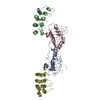 4hrmC  4hrnC 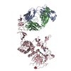 1n8zS  2xeeS S: Starting model for refinement C: citing same article ( |
|---|---|
| Similar structure data |


-Links
 PDBj
PDBj


- Assembly
| Deposited unit | 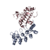
| ||||||||
|---|---|---|---|---|---|---|---|---|---|
| 1 |
| ||||||||
| Unit cell |
|
-Components
| #1: Protein | Mass: 22208.281 Da / Num. of mol.: 1 Fragment: N-terminal extracellular domain I, UNP RESIDUES 24-219 Mutation: N46D, N102D, N165D Source method: isolated from a genetically manipulated source Source: (gene. exp.) Homo sapiens (human) / Gene: ERBB2, HER2, MLN19, NEU, NGL / Plasmid: pFL / Cell line (production host): Sf9 / Production host:   Spodoptera frugiperda (fall armyworm) Spodoptera frugiperda (fall armyworm)References: UniProt: P04626, receptor protein-tyrosine kinase |
|---|---|
| #2: Protein | Mass: 18558.678 Da / Num. of mol.: 1 Source method: isolated from a genetically manipulated source Source: (gene. exp.) synthetic (others) / Plasmid: pQE30 / Production host:  |
| #3: Water | ChemComp-HOH /  Mass: 18.015 Da / Num. of mol.: 31 / Source method: isolated from a natural source / Formula: H2O Mass: 18.015 Da / Num. of mol.: 31 / Source method: isolated from a natural source / Formula: H2O |
| Has protein modification | Y |
-Experimental details
-Experiment
| Experiment | Method: X-RAY DIFFRACTION / Number of used crystals: 1 |
|---|
- Sample preparation
| Crystal | Density Matthews: 2.65 Å3/Da / Density % sol: 53.55 % |
|---|---|
| Crystal grow | Temperature: 293 K / Method: vapor diffusion / pH: 5.6 Details: 0.2M ammonium acetate, 0.1M sodium citrate, 30% PEG 4000, pH 5.6, vapor diffusion, temperature 293K |
-Data collection
| Diffraction | Mean temperature: 90 K | ||||||||||||||||||||||||||||||||||||||||||||||||||||||||||||||||||||||||||||||||||||||||||||||||||||||||||||||||||||||||||||||||||||||||||||||||||||||||||||||||||||||||||||||||||||||||||||||||||||||||||||||||||
|---|---|---|---|---|---|---|---|---|---|---|---|---|---|---|---|---|---|---|---|---|---|---|---|---|---|---|---|---|---|---|---|---|---|---|---|---|---|---|---|---|---|---|---|---|---|---|---|---|---|---|---|---|---|---|---|---|---|---|---|---|---|---|---|---|---|---|---|---|---|---|---|---|---|---|---|---|---|---|---|---|---|---|---|---|---|---|---|---|---|---|---|---|---|---|---|---|---|---|---|---|---|---|---|---|---|---|---|---|---|---|---|---|---|---|---|---|---|---|---|---|---|---|---|---|---|---|---|---|---|---|---|---|---|---|---|---|---|---|---|---|---|---|---|---|---|---|---|---|---|---|---|---|---|---|---|---|---|---|---|---|---|---|---|---|---|---|---|---|---|---|---|---|---|---|---|---|---|---|---|---|---|---|---|---|---|---|---|---|---|---|---|---|---|---|---|---|---|---|---|---|---|---|---|---|---|---|---|---|---|---|---|
| Diffraction source | Source: SYNCHROTRON / Site: SLS  / Beamline: X06SA / Wavelength: 1 Å / Beamline: X06SA / Wavelength: 1 Å | ||||||||||||||||||||||||||||||||||||||||||||||||||||||||||||||||||||||||||||||||||||||||||||||||||||||||||||||||||||||||||||||||||||||||||||||||||||||||||||||||||||||||||||||||||||||||||||||||||||||||||||||||||
| Detector | Type: PSI PILATUS 6M / Detector: PIXEL / Date: Aug 21, 2011 | ||||||||||||||||||||||||||||||||||||||||||||||||||||||||||||||||||||||||||||||||||||||||||||||||||||||||||||||||||||||||||||||||||||||||||||||||||||||||||||||||||||||||||||||||||||||||||||||||||||||||||||||||||
| Radiation | Protocol: SINGLE WAVELENGTH / Monochromatic (M) / Laue (L): M / Scattering type: x-ray | ||||||||||||||||||||||||||||||||||||||||||||||||||||||||||||||||||||||||||||||||||||||||||||||||||||||||||||||||||||||||||||||||||||||||||||||||||||||||||||||||||||||||||||||||||||||||||||||||||||||||||||||||||
| Radiation wavelength | Wavelength: 1 Å / Relative weight: 1 | ||||||||||||||||||||||||||||||||||||||||||||||||||||||||||||||||||||||||||||||||||||||||||||||||||||||||||||||||||||||||||||||||||||||||||||||||||||||||||||||||||||||||||||||||||||||||||||||||||||||||||||||||||
| Reflection | Highest resolution: 2.55 Å / Num. obs: 14533 / % possible obs: 98.7 % / Observed criterion σ(I): -3 / Biso Wilson estimate: 40.398 Å2 / Rmerge F obs: 0.119 / Rmerge(I) obs: 0.071 / Rrim(I) all: 0.083 / Net I/σ(I): 17.83 / Num. measured all: 53167 | ||||||||||||||||||||||||||||||||||||||||||||||||||||||||||||||||||||||||||||||||||||||||||||||||||||||||||||||||||||||||||||||||||||||||||||||||||||||||||||||||||||||||||||||||||||||||||||||||||||||||||||||||||
| Reflection shell | Diffraction-ID: 1
|
-Phasing
| Phasing | Method: molecular replacement | |||||||||
|---|---|---|---|---|---|---|---|---|---|---|
| Phasing MR | Model details: Phaser MODE: MR_AUTO
|
- Processing
| Software |
| ||||||||||||||||||||||||||||||||||||||||||
|---|---|---|---|---|---|---|---|---|---|---|---|---|---|---|---|---|---|---|---|---|---|---|---|---|---|---|---|---|---|---|---|---|---|---|---|---|---|---|---|---|---|---|---|
| Refinement | Method to determine structure: MOLECULAR REPLACEMENT Starting model: 1N8Z, 2XEE Resolution: 2.55→46.817 Å / Occupancy max: 1 / Occupancy min: 0.5 / FOM work R set: 0.8233 / SU ML: 0.31 / σ(F): 1.99 / Phase error: 24.01 / Stereochemistry target values: ML
| ||||||||||||||||||||||||||||||||||||||||||
| Solvent computation | Shrinkage radii: 0.9 Å / VDW probe radii: 1.2 Å / Solvent model: FLAT BULK SOLVENT MODEL | ||||||||||||||||||||||||||||||||||||||||||
| Displacement parameters | Biso max: 77.58 Å2 / Biso mean: 32.9895 Å2 / Biso min: 11.46 Å2 | ||||||||||||||||||||||||||||||||||||||||||
| Refinement step | Cycle: LAST / Resolution: 2.55→46.817 Å
| ||||||||||||||||||||||||||||||||||||||||||
| Refine LS restraints |
| ||||||||||||||||||||||||||||||||||||||||||
| LS refinement shell | Refine-ID: X-RAY DIFFRACTION / Total num. of bins used: 5
|
