 Movie
Movie Controller

[English] 日本語
 Yorodumi
- PDB-5fqn: Crystal structure of M. musculus protein arginine methyltransfera... -
+ Open data

- Basic information
| Entry | Database: PDB / ID: 5fqn | ||||||
|---|---|---|---|---|---|---|---|
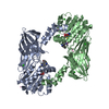
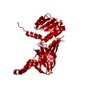
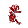
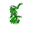
| Title | Crystal structure of M. musculus protein arginine methyltransferase PRMT6 with SAH at 1.65 Angstroms | ||||||
 Components Components | PROTEIN ARGININE METHYLTRANSFERASE 6 | ||||||
 Keywords Keywords | TRANSFERASE / S-ADENOSYL-L-METHIONINE | ||||||
| Function / homology |  Function and homology information Function and homology informationhistone H2AR3 methyltransferase activity / protein-arginine omega-N monomethyltransferase activity / RUNX1 regulates genes involved in megakaryocyte differentiation and platelet function / histone H3R2 methyltransferase activity / RMTs methylate histone arginines / type I protein arginine methyltransferase / protein-arginine omega-N asymmetric methyltransferase activity / histone H4R3 methyltransferase activity / protein-arginine N-methyltransferase activity / regulation of mitochondrion organization ...histone H2AR3 methyltransferase activity / protein-arginine omega-N monomethyltransferase activity / RUNX1 regulates genes involved in megakaryocyte differentiation and platelet function / histone H3R2 methyltransferase activity / RMTs methylate histone arginines / type I protein arginine methyltransferase / protein-arginine omega-N asymmetric methyltransferase activity / histone H4R3 methyltransferase activity / protein-arginine N-methyltransferase activity / regulation of mitochondrion organization / histone H3 methyltransferase activity / histone methyltransferase activity / negative regulation of ubiquitin-dependent protein catabolic process / regulation of signal transduction by p53 class mediator / cellular senescence / methylation / histone binding / chromatin remodeling / DNA repair / negative regulation of DNA-templated transcription / chromatin binding / regulation of DNA-templated transcription / nucleolus / negative regulation of transcription by RNA polymerase II / nucleoplasm / nucleus Similarity search - Function | ||||||
| Biological species |  | ||||||
| Method |  X-RAY DIFFRACTION / SYNCHROTRON / MOLECULAR REPLACEMENT / Resolution: 1.657 Å X-RAY DIFFRACTION / SYNCHROTRON / MOLECULAR REPLACEMENT / Resolution: 1.657 Å | ||||||
 Authors Authors | Bonnefond, L. / Cavarelli, J. | ||||||
 Citation Citation | Journal: To be Published Title: Crystal Strcutures of Prmt6 in Complex with Sah in Alternative Conformations Authors: Bonnefond, L. / Cura, V. / Troffer-Charlier, N. / Cavarelli, J. | ||||||
| History |
|
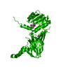
- Structure visualization
| Structure viewer | Molecule: MolmilJmol/JSmol |
|---|
- Downloads & links
-Download
| PDBx/mmCIF format | 5fqn.cif.gz | 84.3 KB | Display | PDBx/mmCIF format |
|---|---|---|---|---|
| PDB format | pdb5fqn.ent.gz | 61.6 KB | Display | PDB format |
| PDBx/mmJSON format | 5fqn.json.gz | Tree view | PDBx/mmJSON format | |
| Others |  Other downloads Other downloads |
-Validation report
| Arichive directory | https://data.pdbj.org/pub/pdb/validation_reports/fq/5fqnftp://data.pdbj.org/pub/pdb/validation_reports/fq/5fqn | HTTPS FTP |
|---|
-Related structure data
| Related structure data | 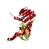 5fqoC 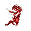 4c08S C: citing same article ( S: Starting model for refinement |
|---|---|
| Similar structure data |


-Links
 PDBj
PDBj
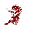
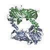
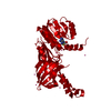
- Assembly
| Deposited unit | 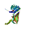
| ||||||||
|---|---|---|---|---|---|---|---|---|---|
| 1 | 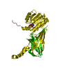
| ||||||||
| Unit cell |
|
-Components
| #1: Protein | Mass: 44355.965 Da / Num. of mol.: 1 Source method: isolated from a genetically manipulated source Source: (gene. exp.)  References: UniProt: Q6NZB1, Transferases; Transferring one-carbon groups; Methyltransferases, EC: 2.1.1.125 |
|---|---|
| #2: Chemical | ChemComp-SAH /   Mass: 384.411 Da / Num. of mol.: 1 / Source method: obtained synthetically / Formula: C14H20N6O5S Mass: 384.411 Da / Num. of mol.: 1 / Source method: obtained synthetically / Formula: C14H20N6O5S |
| #3: Water | ChemComp-HOH /  Mass: 18.015 Da / Num. of mol.: 178 / Source method: isolated from a natural source / Formula: H2O Mass: 18.015 Da / Num. of mol.: 178 / Source method: isolated from a natural source / Formula: H2O |
| Has protein modification | Y |
| Sequence details | L315P NATURAL VARIANT |
-Experimental details
-Experiment
| Experiment | Method: X-RAY DIFFRACTION / Number of used crystals: 1 |
|---|
- Sample preparation
| Crystal | Density Matthews: 2.11 Å3/Da / Density % sol: 41.65 % / Description: NONE |
|---|---|
| Crystal grow | pH: 8 / Details: 100 MM TRIS PH 8.0 200 MM MGCL2 12% PEG 6000 |
-Data collection
| Diffraction | Mean temperature: 100 K |
|---|---|
| Diffraction source | Source: SYNCHROTRON / Site: SOLEIL  / Beamline: PROXIMA 2 / Wavelength: 0.9801 / Beamline: PROXIMA 2 / Wavelength: 0.9801 |
| Detector | Type: ADSC QUANTUM 315 / Detector: CCD / Date: Mar 26, 2015 Details: A CONVEX PREFOCUSSING MIRROR AND A KIRKPATRICK- BAEZ PAIR OF FOCUSSING MIRRORS |
| Radiation | Monochromator: CRYOGENICALLY COOLED CHANNEL CUT CRYSTAL MONOCHROMATOR Protocol: SINGLE WAVELENGTH / Monochromatic (M) / Laue (L): M / Scattering type: x-ray |
| Radiation wavelength | Wavelength: 0.9801 Å / Relative weight: 1 |
| Reflection | Resolution: 1.65→40.8 Å / Num. obs: 43482 / % possible obs: 99.9 % / Observed criterion σ(I): 1 / Redundancy: 5.6 % / Biso Wilson estimate: 23.97 Å2 / Rmerge(I) obs: 0.08 / Net I/σ(I): 10.1 |
| Reflection shell | Resolution: 1.66→1.69 Å / Redundancy: 5.6 % / Rmerge(I) obs: 1.724 / Mean I/σ(I) obs: 0.8 / % possible all: 99 |
- Processing
| Software |
| |||||||||||||||||||||||||||||||||||||||||||||||||||||||||||||||||||||||||||||||||||||||||||||||||||||||||||||||||||||||
|---|---|---|---|---|---|---|---|---|---|---|---|---|---|---|---|---|---|---|---|---|---|---|---|---|---|---|---|---|---|---|---|---|---|---|---|---|---|---|---|---|---|---|---|---|---|---|---|---|---|---|---|---|---|---|---|---|---|---|---|---|---|---|---|---|---|---|---|---|---|---|---|---|---|---|---|---|---|---|---|---|---|---|---|---|---|---|---|---|---|---|---|---|---|---|---|---|---|---|---|---|---|---|---|---|---|---|---|---|---|---|---|---|---|---|---|---|---|---|---|---|
| Refinement | Method to determine structure: MOLECULAR REPLACEMENT Starting model: PDB ENTRY 4C08 Resolution: 1.657→40.772 Å / SU ML: 0.2 / σ(F): 1.36 / Phase error: 23.61 / Stereochemistry target values: ML
| |||||||||||||||||||||||||||||||||||||||||||||||||||||||||||||||||||||||||||||||||||||||||||||||||||||||||||||||||||||||
| Solvent computation | Shrinkage radii: 0.9 Å / VDW probe radii: 1.11 Å / Solvent model: FLAT BULK SOLVENT MODEL | |||||||||||||||||||||||||||||||||||||||||||||||||||||||||||||||||||||||||||||||||||||||||||||||||||||||||||||||||||||||
| Displacement parameters | Biso mean: 30.2 Å2 | |||||||||||||||||||||||||||||||||||||||||||||||||||||||||||||||||||||||||||||||||||||||||||||||||||||||||||||||||||||||
| Refinement step | Cycle: LAST / Resolution: 1.657→40.772 Å
| |||||||||||||||||||||||||||||||||||||||||||||||||||||||||||||||||||||||||||||||||||||||||||||||||||||||||||||||||||||||
| Refine LS restraints |
| |||||||||||||||||||||||||||||||||||||||||||||||||||||||||||||||||||||||||||||||||||||||||||||||||||||||||||||||||||||||
| LS refinement shell |
|
