 Movie
Movie Controller


+ Open data

- Basic information
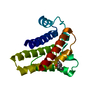
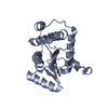
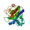
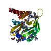
| Entry | Database: PDB / ID: 4h79 | ||||||
|---|---|---|---|---|---|---|---|
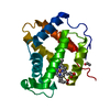
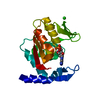
| Title | Crystal structure of CasB from Thermobifida fusca | ||||||
 Components Components | CRISPR-associated protein, Cse2 family | ||||||
 Keywords Keywords | DNA BINDING PROTEIN / CRISPR / Cascade / CasB / CRISPR-assoicated protein / Nucleic acid binding protein | ||||||
| Function / homology | CRISPR-associated protein Cse2 / CRISPR-associated protein Cse2 / Cse2 superfamily / CRISPR-associated protein Cse2 (CRISPR_cse2) / Peroxidase; domain 1 / Orthogonal Bundle / Mainly Alpha / CRISPR-associated protein, Cse2 family Function and homology information Function and homology information | ||||||
| Biological species |   Thermobifida fusca (bacteria) Thermobifida fusca (bacteria) | ||||||
| Method |  X-RAY DIFFRACTION / SYNCHROTRON / MOLECULAR REPLACEMENT / Resolution: 1.9 Å X-RAY DIFFRACTION / SYNCHROTRON / MOLECULAR REPLACEMENT / Resolution: 1.9 Å | ||||||
 Authors Authors | Ke, A. / Nam, K.H. | ||||||
 Citation Citation | Journal: Febs Lett. / Year: 2012 Title: Nucleic acid binding surface and dimer interface revealed by CRISPR-associated CasB protein structures. Authors: Nam, K.H. / Huang, Q. / Ke, A. | ||||||
| History |
|
- Structure visualization
| Structure viewer | Molecule: MolmilJmol/JSmol |
|---|
- Downloads & links
-Download
| PDBx/mmCIF format | 4h79.cif.gz | 90.6 KB | Display | PDBx/mmCIF format |
|---|---|---|---|---|
| PDB format | pdb4h79.ent.gz | 69.1 KB | Display | PDB format |
| PDBx/mmJSON format | 4h79.json.gz | Tree view | PDBx/mmJSON format | |
| Others |  Other downloads Other downloads |
-Validation report
| Arichive directory | https://data.pdbj.org/pub/pdb/validation_reports/h7/4h79ftp://data.pdbj.org/pub/pdb/validation_reports/h7/4h79 | HTTPS FTP |
|---|
-Related structure data
| Related structure data | 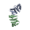 4h7aC 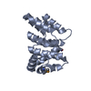 2zcaS S: Starting model for refinement C: citing same article ( |
|---|---|
| Similar structure data |


-Links
 PDBj
PDBj
- Assembly
| Deposited unit | 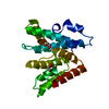
| ||||||||
|---|---|---|---|---|---|---|---|---|---|
| 1 |
| ||||||||
| Unit cell |
|
-Components
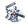
| #1: Protein | Mass: 24682.883 Da / Num. of mol.: 1 Source method: isolated from a genetically manipulated source Source: (gene. exp.) Thermobifida fusca (bacteria) / Strain: YX / Gene: Tfu_1599 / Plasmid: modified pSUMO / Production host: | ||
|---|---|---|---|
| #2: Chemical |   Mass: 62.068 Da / Num. of mol.: 2 / Source method: obtained synthetically / Formula: C2H6O2 Mass: 62.068 Da / Num. of mol.: 2 / Source method: obtained synthetically / Formula: C2H6O2#3: Water | ChemComp-HOH / |  Mass: 18.015 Da / Num. of mol.: 89 / Source method: isolated from a natural source / Formula: H2O Mass: 18.015 Da / Num. of mol.: 89 / Source method: isolated from a natural source / Formula: H2O |
-Experimental details
-Experiment
| Experiment | Method: X-RAY DIFFRACTION / Number of used crystals: 1 |
|---|
- Sample preparation
| Crystal | Density Matthews: 3.08 Å3/Da / Density % sol: 60.08 % |
|---|---|
| Crystal grow | Temperature: 291.5 K / Method: vapor diffusion, hanging drop / pH: 8 Details: 0.1M Tris-HCl, pH 8.0-8.5, 2% (v/v) ethanol, VAPOR DIFFUSION, HANGING DROP, temperature 291.5K |
-Data collection
| Diffraction | Mean temperature: 100 K |
|---|---|
| Diffraction source | Source: SYNCHROTRON / Site: CHESS  / Beamline: A1 / Wavelength: 0.977 Å / Beamline: A1 / Wavelength: 0.977 Å |
| Detector | Type: ADSC QUANTUM 210 / Detector: CCD / Date: Nov 18, 2010 |
| Radiation | Monochromator: Si 111 CHANNEL / Protocol: SINGLE WAVELENGTH / Monochromatic (M) / Laue (L): M / Scattering type: x-ray |
| Radiation wavelength | Wavelength: 0.977 Å / Relative weight: 1 |
| Reflection | Resolution: 1.9→20 Å / Num. obs: 22861 / % possible obs: 96.3 % / Observed criterion σ(F): 2 / Observed criterion σ(I): 2 |
| Reflection shell | Resolution: 1.9→1.93 Å / % possible all: 78.1 |
- Processing
| Software |
| ||||||||||||||||||||||||||||||||||||||||||||||||||||||||||||
|---|---|---|---|---|---|---|---|---|---|---|---|---|---|---|---|---|---|---|---|---|---|---|---|---|---|---|---|---|---|---|---|---|---|---|---|---|---|---|---|---|---|---|---|---|---|---|---|---|---|---|---|---|---|---|---|---|---|---|---|---|---|
| Refinement | Method to determine structure: MOLECULAR REPLACEMENT Starting model: PDB ENTRY 2ZCA Resolution: 1.9→20 Å / Cor.coef. Fo:Fc: 0.973 / Cor.coef. Fo:Fc free: 0.955 / Occupancy max: 1 / Occupancy min: 1 / SU B: 5.507 / SU ML: 0.071 / Cross valid method: THROUGHOUT / σ(F): 0 / ESU R: 0.128 / ESU R Free: 0.102 / Stereochemistry target values: MAXIMUM LIKELIHOOD Details: HYDROGENS HAVE BEEN USED IF PRESENT IN THE INPUT U VALUES : RESIDUAL ONLY
| ||||||||||||||||||||||||||||||||||||||||||||||||||||||||||||
| Solvent computation | Ion probe radii: 0.8 Å / Shrinkage radii: 0.8 Å / VDW probe radii: 1.2 Å / Solvent model: MASK | ||||||||||||||||||||||||||||||||||||||||||||||||||||||||||||
| Displacement parameters | Biso max: 121.48 Å2 / Biso mean: 37.0509 Å2 / Biso min: 14.82 Å2
| ||||||||||||||||||||||||||||||||||||||||||||||||||||||||||||
| Refinement step | Cycle: LAST / Resolution: 1.9→20 Å
| ||||||||||||||||||||||||||||||||||||||||||||||||||||||||||||
| Refine LS restraints |
| ||||||||||||||||||||||||||||||||||||||||||||||||||||||||||||
| LS refinement shell | Resolution: 1.9→1.949 Å / Total num. of bins used: 20
| ||||||||||||||||||||||||||||||||||||||||||||||||||||||||||||
| Refinement TLS params. | Method: refined / Origin x: -29.2936 Å / Origin y: 32.5466 Å / Origin z: 9.8554 Å
|
