 Movie
Movie Controller

[English] 日本語
 Yorodumi
- PDB-2fcl: Crystal structure of a putative nucleotidyltransferase (tm1012) f... -
+ Open data

- Basic information
| Entry | Database: PDB / ID: 2fcl | ||||||
|---|---|---|---|---|---|---|---|
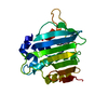
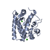
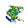
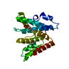
| Title | Crystal structure of a putative nucleotidyltransferase (tm1012) from Thermotoga maritima at 1.35 A resolution | ||||||
 Components Components | hypothetical protein TM1012 | ||||||
 Keywords Keywords | TRANSFERASE / PUTATIVE NUCLEOTIDYLTRANSFERASE / STRUCTURAL GENOMICS / JOINT CENTER FOR STRUCTURAL GENOMICS / JCSG / PROTEIN STRUCTURE INITIATIVE / PSI-2 | ||||||
| Function / homology |  Function and homology information Function and homology informationAminoglycoside-2''-adenylyltransferase / Aminoglycoside-2''-adenylyltransferase / Beta Polymerase; domain 2 - #40 / Beta Polymerase; domain 2 / Nucleotidyltransferase superfamily / 2-Layer Sandwich / Alpha Beta Similarity search - Domain/homology | ||||||
| Biological species |   Thermotoga maritima (bacteria) Thermotoga maritima (bacteria) | ||||||
| Method |  X-RAY DIFFRACTION / SYNCHROTRON / MOLECULAR REPLACEMENT / molecular replacement / Resolution: 1.2 Å X-RAY DIFFRACTION / SYNCHROTRON / MOLECULAR REPLACEMENT / molecular replacement / Resolution: 1.2 Å | ||||||
 Authors Authors | Joint Center for Structural Genomics (JCSG) | ||||||
 Citation Citation | Journal: To be published Title: Crystal structure of hypothetical protein (tm1012) from Thermotoga maritima at 1.35 A resolution Authors: Joint Center for Structural Genomics (JCSG) | ||||||
| History |
| ||||||
| Remark 999 | SEQUENCE THE PROTEIN WAS REDUCTIVELY METHYLATED PRIOR TO CRYSTALLIZATION. |
- Structure visualization
| Structure viewer | Molecule: MolmilJmol/JSmol |
|---|
- Downloads & links
-Download
| PDBx/mmCIF format | 2fcl.cif.gz | 89.2 KB | Display | PDBx/mmCIF format |
|---|---|---|---|---|
| PDB format | pdb2fcl.ent.gz | 66.2 KB | Display | PDB format |
| PDBx/mmJSON format | 2fcl.json.gz | Tree view | PDBx/mmJSON format | |
| Others |  Other downloads Other downloads |
-Validation report
| Arichive directory | https://data.pdbj.org/pub/pdb/validation_reports/fc/2fclftp://data.pdbj.org/pub/pdb/validation_reports/fc/2fcl | HTTPS FTP |
|---|
-Related structure data
| Related structure data | 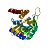 2ewrS S: Starting model for refinement |
|---|---|
| Similar structure data | |
| Other databases |


-Links
 PDBj
PDBj


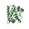
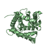
- Assembly
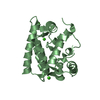
| Deposited unit | 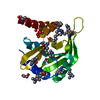
| ||||||||
|---|---|---|---|---|---|---|---|---|---|
| 1 |
| ||||||||
| Unit cell |
|
-Components
| #1: Protein | Mass: 20493.795 Da / Num. of mol.: 1 Source method: isolated from a genetically manipulated source Source: (gene. exp.) Thermotoga maritima (bacteria) / Gene: tm1012 / Production host: | ||
|---|---|---|---|
| #2: Chemical | ChemComp-UNL / Num. of mol.: 1 / Source method: obtained synthetically | ||
| #3: Chemical |   Mass: 62.068 Da / Num. of mol.: 2 / Source method: obtained synthetically / Formula: C2H6O2 Mass: 62.068 Da / Num. of mol.: 2 / Source method: obtained synthetically / Formula: C2H6O2#4: Water | ChemComp-HOH / |  Mass: 18.015 Da / Num. of mol.: 261 / Source method: isolated from a natural source / Formula: H2O Mass: 18.015 Da / Num. of mol.: 261 / Source method: isolated from a natural source / Formula: H2O |
-Experimental details
-Experiment
| Experiment | Method: X-RAY DIFFRACTION / Number of used crystals: 1 |
|---|
- Sample preparation
| Crystal | Density Matthews: 2.18 Å3/Da / Density % sol: 43.55 % |
|---|---|
| Crystal grow | Temperature: 277 K / Method: vapor diffusion, sitting drop, nanodrop / pH: 5.6 Details: 15.0% Glycerol, 0.17M NH4OAc, 25.5% PEG-4000, 0.1M Citrate, pH 5.6, VAPOR DIFFUSION, SITTING DROP, NANODROP, temperature 277K |
-Data collection
| Diffraction | Mean temperature: 100 K | ||||||||||||||||||||||||||||||||||||||||||||||||||||||||||||||||||||||||||||||||||||||||||||||||||||||||||||||||||||||||||||||||||||||||||||||||||||||||||||||||||||||||
|---|---|---|---|---|---|---|---|---|---|---|---|---|---|---|---|---|---|---|---|---|---|---|---|---|---|---|---|---|---|---|---|---|---|---|---|---|---|---|---|---|---|---|---|---|---|---|---|---|---|---|---|---|---|---|---|---|---|---|---|---|---|---|---|---|---|---|---|---|---|---|---|---|---|---|---|---|---|---|---|---|---|---|---|---|---|---|---|---|---|---|---|---|---|---|---|---|---|---|---|---|---|---|---|---|---|---|---|---|---|---|---|---|---|---|---|---|---|---|---|---|---|---|---|---|---|---|---|---|---|---|---|---|---|---|---|---|---|---|---|---|---|---|---|---|---|---|---|---|---|---|---|---|---|---|---|---|---|---|---|---|---|---|---|---|---|---|---|---|---|
| Diffraction source | Source: SYNCHROTRON / Site: SSRL  / Beamline: BL11-1 / Wavelength: 0.979454 / Wavelength: 0.979454 Å / Beamline: BL11-1 / Wavelength: 0.979454 / Wavelength: 0.979454 Å | ||||||||||||||||||||||||||||||||||||||||||||||||||||||||||||||||||||||||||||||||||||||||||||||||||||||||||||||||||||||||||||||||||||||||||||||||||||||||||||||||||||||||
| Detector | Type: ADSC QUANTUM 315 / Detector: CCD / Date: Apr 27, 2005 / Details: Flat mirror (vertical focusing) | ||||||||||||||||||||||||||||||||||||||||||||||||||||||||||||||||||||||||||||||||||||||||||||||||||||||||||||||||||||||||||||||||||||||||||||||||||||||||||||||||||||||||
| Radiation | Monochromator: Single crystal Si(111) bent monochromator (horizontal focusing) Protocol: SINGLE WAVELENGTH / Monochromatic (M) / Laue (L): M / Scattering type: x-ray | ||||||||||||||||||||||||||||||||||||||||||||||||||||||||||||||||||||||||||||||||||||||||||||||||||||||||||||||||||||||||||||||||||||||||||||||||||||||||||||||||||||||||
| Radiation wavelength | Wavelength: 0.979454 Å / Relative weight: 1 | ||||||||||||||||||||||||||||||||||||||||||||||||||||||||||||||||||||||||||||||||||||||||||||||||||||||||||||||||||||||||||||||||||||||||||||||||||||||||||||||||||||||||
| Reflection | Resolution: 1.2→25.88 Å / Num. obs: 39827 / % possible obs: 73.5 % / Redundancy: 2.3 % / Biso Wilson estimate: 11.7 Å2 / Rmerge(I) obs: 0.034 / Rsym value: 0.034 / Net I/σ(I): 20.3 | ||||||||||||||||||||||||||||||||||||||||||||||||||||||||||||||||||||||||||||||||||||||||||||||||||||||||||||||||||||||||||||||||||||||||||||||||||||||||||||||||||||||||
| Reflection shell |
|
-Phasing
| Phasing | Method: molecular replacement |
|---|
- Processing
| Software |
| |||||||||||||||||||||||||||||||||||||||||||||||||||||||||||||||||||||||||||||||||||||||||||||||||||||||||||||||||||||||||||||||||||||||
|---|---|---|---|---|---|---|---|---|---|---|---|---|---|---|---|---|---|---|---|---|---|---|---|---|---|---|---|---|---|---|---|---|---|---|---|---|---|---|---|---|---|---|---|---|---|---|---|---|---|---|---|---|---|---|---|---|---|---|---|---|---|---|---|---|---|---|---|---|---|---|---|---|---|---|---|---|---|---|---|---|---|---|---|---|---|---|---|---|---|---|---|---|---|---|---|---|---|---|---|---|---|---|---|---|---|---|---|---|---|---|---|---|---|---|---|---|---|---|---|---|---|---|---|---|---|---|---|---|---|---|---|---|---|---|---|---|
| Refinement | Method to determine structure: MOLECULAR REPLACEMENT Starting model: pdb entry 2ewr Resolution: 1.2→24.28 Å / Cor.coef. Fo:Fc: 0.976 / Cor.coef. Fo:Fc free: 0.962 / SU B: 0.958 / SU ML: 0.022 / Cross valid method: THROUGHOUT / ESU R: 0.049 / ESU R Free: 0.048 / Stereochemistry target values: MAXIMUM LIKELIHOOD Details: (1) HYDROGENS HAVE BEEN ADDED IN THE RIDING POSITIONS. (2) THE NOMINAL RESOLUTION IS 1.35 A WITH 4948 OBSERVED REFLECTIONS BETWEEN 1.35-1.20 (33.6% COMPLETE FOR THIS SHELL) INCLUDED IN THE ...Details: (1) HYDROGENS HAVE BEEN ADDED IN THE RIDING POSITIONS. (2) THE NOMINAL RESOLUTION IS 1.35 A WITH 4948 OBSERVED REFLECTIONS BETWEEN 1.35-1.20 (33.6% COMPLETE FOR THIS SHELL) INCLUDED IN THE REFINEMENT. (3) DENSITY NEAR RESIDUES 116 AND 129 WAS MODELED AS AN UNKNOWN LIGAND (UNL).
| |||||||||||||||||||||||||||||||||||||||||||||||||||||||||||||||||||||||||||||||||||||||||||||||||||||||||||||||||||||||||||||||||||||||
| Solvent computation | Ion probe radii: 0.8 Å / Shrinkage radii: 0.8 Å / VDW probe radii: 1.2 Å / Solvent model: MASK | |||||||||||||||||||||||||||||||||||||||||||||||||||||||||||||||||||||||||||||||||||||||||||||||||||||||||||||||||||||||||||||||||||||||
| Displacement parameters | Biso mean: 13.634 Å2
| |||||||||||||||||||||||||||||||||||||||||||||||||||||||||||||||||||||||||||||||||||||||||||||||||||||||||||||||||||||||||||||||||||||||
| Refinement step | Cycle: LAST / Resolution: 1.2→24.28 Å
| |||||||||||||||||||||||||||||||||||||||||||||||||||||||||||||||||||||||||||||||||||||||||||||||||||||||||||||||||||||||||||||||||||||||
| Refine LS restraints |
| |||||||||||||||||||||||||||||||||||||||||||||||||||||||||||||||||||||||||||||||||||||||||||||||||||||||||||||||||||||||||||||||||||||||
| LS refinement shell | Resolution: 1.201→1.232 Å / Total num. of bins used: 20
|
