 Movie
Movie Controller


+ Open data

- Basic information
| Entry | Database: PDB / ID: 6ajr | ||||||
|---|---|---|---|---|---|---|---|
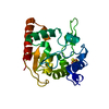
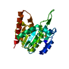
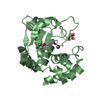
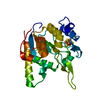
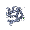
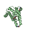
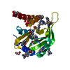
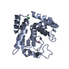
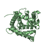
| Title | Complex form of Uracil DNA glycosylase X and uracil | ||||||
 Components Components | Uracil DNA glycosylase superfamily protein | ||||||
 Keywords Keywords | DNA BINDING PROTEIN / DNA repair / Base excision / DNA-Protein crosslink. | ||||||
| Function / homology |  Function and homology information Function and homology informationdeaminated base DNA N-glycosylase activity / 4 iron, 4 sulfur cluster binding / DNA repair / metal ion binding Similarity search - Function | ||||||
| Biological species |  Mycobacterium smegmatis MC2 155 (bacteria) Mycobacterium smegmatis MC2 155 (bacteria) | ||||||
| Method |  X-RAY DIFFRACTION / SYNCHROTRON / MIR / Resolution: 1.341 Å X-RAY DIFFRACTION / SYNCHROTRON / MIR / Resolution: 1.341 Å | ||||||
 Authors Authors | Ahn, W.C. / Aroli, S. / Varshney, U. / Woo, E.J. | ||||||
| Funding support |  Korea, Republic Of, 1items Korea, Republic Of, 1items
| ||||||
 Citation Citation | Journal: Nat.Chem.Biol. / Year: 2019 Title: Covalent binding of uracil DNA glycosylase UdgX to abasic DNA upon uracil excision. Authors: Ahn, W.C. / Aroli, S. / Kim, J.H. / Moon, J.H. / Lee, G.S. / Lee, M.H. / Sang, P.B. / Oh, B.H. / Varshney, U. / Woo, E.J. | ||||||
| History |
|
- Structure visualization
| Structure viewer | Molecule: MolmilJmol/JSmol |
|---|
- Downloads & links
-Download
| PDBx/mmCIF format | 6ajr.cif.gz | 97.6 KB | Display | PDBx/mmCIF format |
|---|---|---|---|---|
| PDB format | pdb6ajr.ent.gz | 72.4 KB | Display | PDB format |
| PDBx/mmJSON format | 6ajr.json.gz | Tree view | PDBx/mmJSON format | |
| Others |  Other downloads Other downloads |
-Validation report
| Arichive directory | https://data.pdbj.org/pub/pdb/validation_reports/aj/6ajrftp://data.pdbj.org/pub/pdb/validation_reports/aj/6ajr | HTTPS FTP |
|---|
-Related structure data


-Links
 PDBj
PDBj- Assembly
| Deposited unit | 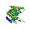
| ||||||||
|---|---|---|---|---|---|---|---|---|---|
| 1 |
| ||||||||
| Unit cell |
|
-Components
| #1: Protein | Mass: 22201.283 Da / Num. of mol.: 1 Source method: isolated from a genetically manipulated source Source: (gene. exp.) Mycobacterium smegmatis MC2 155 (bacteria)Strain: MC2 155 / Gene: MSMEG_0265 / Production host: |
|---|---|
| #2: Chemical | ChemComp-SF4 /   Mass: 351.640 Da / Num. of mol.: 1 / Source method: obtained synthetically / Formula: Fe4S4 Mass: 351.640 Da / Num. of mol.: 1 / Source method: obtained synthetically / Formula: Fe4S4 |
| #3: Chemical | ChemComp-URA /   Mass: 112.087 Da / Num. of mol.: 1 / Source method: obtained synthetically / Formula: C4H4N2O2 / Feature type: SUBJECT OF INVESTIGATION Mass: 112.087 Da / Num. of mol.: 1 / Source method: obtained synthetically / Formula: C4H4N2O2 / Feature type: SUBJECT OF INVESTIGATION |
| #4: Water | ChemComp-HOH /  Mass: 18.015 Da / Num. of mol.: 299 / Source method: isolated from a natural source / Formula: H2O Mass: 18.015 Da / Num. of mol.: 299 / Source method: isolated from a natural source / Formula: H2O |
-Experimental details
-Experiment
| Experiment | Method: X-RAY DIFFRACTION / Number of used crystals: 1 |
|---|
- Sample preparation
| Crystal | Density Matthews: 2.26 Å3/Da / Density % sol: 45.68 % |
|---|---|
| Crystal grow | Temperature: 290 K / Method: vapor diffusion, sitting drop Details: 2.0M ammonium citrate tribasic pH7.0, 0.1M Bis-Tris propane pH7.0 |
-Data collection
| Diffraction | Mean temperature: 190 K |
|---|---|
| Diffraction source | Source: SYNCHROTRON / Site: PAL/PLS / Beamline: 7A (6B, 6C1) / Wavelength: 0.987 Å |
| Detector | Type: ADSC QUANTUM 270 / Detector: CCD / Date: Jul 13, 2015 |
| Radiation | Protocol: SINGLE WAVELENGTH / Monochromatic (M) / Laue (L): M / Scattering type: x-ray |
| Radiation wavelength | Wavelength: 0.987 Å / Relative weight: 1 |
| Reflection | Resolution: 1.34→50 Å / Num. obs: 43403 / % possible obs: 97.7 % / Redundancy: 6.9 % / Rmerge(I) obs: 0.054 / Net I/σ(I): 54.35 |
| Reflection shell | Resolution: 1.34→1.36 Å / Rmerge(I) obs: 0.238 / Num. unique obs: 2162 |
- Processing
| Software |
| |||||||||||||||||||||||||||||||||||||||||||||||||||||||||||||||||||||||||||||||||||||||||||||||||||||||||
|---|---|---|---|---|---|---|---|---|---|---|---|---|---|---|---|---|---|---|---|---|---|---|---|---|---|---|---|---|---|---|---|---|---|---|---|---|---|---|---|---|---|---|---|---|---|---|---|---|---|---|---|---|---|---|---|---|---|---|---|---|---|---|---|---|---|---|---|---|---|---|---|---|---|---|---|---|---|---|---|---|---|---|---|---|---|---|---|---|---|---|---|---|---|---|---|---|---|---|---|---|---|---|---|---|---|---|
| Refinement | Method to determine structure: MIR / Resolution: 1.341→19.008 Å / SU ML: 0.09 / Cross valid method: FREE R-VALUE / σ(F): 1.34 / Phase error: 14.7
| |||||||||||||||||||||||||||||||||||||||||||||||||||||||||||||||||||||||||||||||||||||||||||||||||||||||||
| Solvent computation | Shrinkage radii: 0.9 Å / VDW probe radii: 1.11 Å | |||||||||||||||||||||||||||||||||||||||||||||||||||||||||||||||||||||||||||||||||||||||||||||||||||||||||
| Refinement step | Cycle: LAST / Resolution: 1.341→19.008 Å
| |||||||||||||||||||||||||||||||||||||||||||||||||||||||||||||||||||||||||||||||||||||||||||||||||||||||||
| Refine LS restraints |
| |||||||||||||||||||||||||||||||||||||||||||||||||||||||||||||||||||||||||||||||||||||||||||||||||||||||||
| LS refinement shell |
|
