 Movie
Movie Controller

[English] 日本語
 Yorodumi
- PDB-4h3t: Crystal structure of CRISPR-associated protein Cse1 from Acidimic... -
+ Open data

- Basic information
| Entry | Database: PDB / ID: 4h3t | ||||||
|---|---|---|---|---|---|---|---|
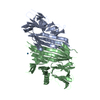
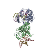
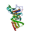
| Title | Crystal structure of CRISPR-associated protein Cse1 from Acidimicrobium ferrooxidans | ||||||
 Components Components | CRISPR-associated protein, Cse1 family | ||||||
 Keywords Keywords | CELL INVASION / Structural Genomics / PSI-Biology / Midwest Center for Structural Genomics / MCSG / CASCADE | ||||||
| Function / homology | Topoisomerase I; Chain A, domain 4 - #100 / CRISPR-associated protein Cse1 / CRISPR-associated protein Cse1 (CRISPR_cse1) / Topoisomerase I; Chain A, domain 4 / Orthogonal Bundle / Mainly Alpha / CRISPR-associated protein, Cse1 family Function and homology information Function and homology information | ||||||
| Biological species |   Acidimicrobium ferrooxidans (bacteria) Acidimicrobium ferrooxidans (bacteria) | ||||||
| Method |  X-RAY DIFFRACTION / SYNCHROTRON / SAD / Resolution: 2.03 Å X-RAY DIFFRACTION / SYNCHROTRON / SAD / Resolution: 2.03 Å | ||||||
 Authors Authors | Michalska, K. / Stols, L. / Clancy, S. / Joachimiak, A. / Midwest Center for Structural Genomics (MCSG) | ||||||
 Citation Citation | Journal: To be Published Title: Crystal structure of CRISPR-associated protein Cse1 from Acidimicrobium ferrooxidans Authors: Michalska, K. / Stols, L. / Clancy, S. / Joachimiak, A. | ||||||
| History |
|
- Structure visualization
| Structure viewer | Molecule: MolmilJmol/JSmol |
|---|
- Downloads & links
-Download
| PDBx/mmCIF format | 4h3t.cif.gz | 225.2 KB | Display | PDBx/mmCIF format |
|---|---|---|---|---|
| PDB format | pdb4h3t.ent.gz | 181.2 KB | Display | PDB format |
| PDBx/mmJSON format | 4h3t.json.gz | Tree view | PDBx/mmJSON format | |
| Others |  Other downloads Other downloads |
-Validation report
| Arichive directory | https://data.pdbj.org/pub/pdb/validation_reports/h3/4h3tftp://data.pdbj.org/pub/pdb/validation_reports/h3/4h3t | HTTPS FTP |
|---|
-Related structure data
| Similar structure data | |
|---|---|
| Other databases |

-Links
 PDBj
PDBj
- Assembly
| Deposited unit | 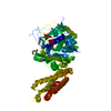
| ||||||||
|---|---|---|---|---|---|---|---|---|---|
| 1 |
| ||||||||
| Unit cell |
| ||||||||
| Components on special symmetry positions |
|
-Components
| #1: Protein | Mass: 60410.285 Da / Num. of mol.: 1 / Mutation: L456I Source method: isolated from a genetically manipulated source Source: (gene. exp.) Acidimicrobium ferrooxidans (bacteria) / Strain: DSM 10331 / Gene: Afer_0988 / Plasmid: pMCSG48 / Production host: | ||||
|---|---|---|---|---|---|
| #2: Chemical |   Mass: 92.094 Da / Num. of mol.: 2 / Source method: obtained synthetically / Formula: C3H8O3 Mass: 92.094 Da / Num. of mol.: 2 / Source method: obtained synthetically / Formula: C3H8O3#3: Water | ChemComp-HOH / |  Mass: 18.015 Da / Num. of mol.: 269 / Source method: isolated from a natural source / Formula: H2O Mass: 18.015 Da / Num. of mol.: 269 / Source method: isolated from a natural source / Formula: H2OHas protein modification | Y | |
-Experimental details
-Experiment
| Experiment | Method: X-RAY DIFFRACTION / Number of used crystals: 1 |
|---|
- Sample preparation
| Crystal | Density Matthews: 3.95 Å3/Da / Density % sol: 68.89 % |
|---|---|
| Crystal grow | Temperature: 289 K / Method: vapor diffusion, sitting drop / pH: 7.5 Details: 0.2 M proline, 0.1 M HEPES/NaOH, 10% PEG3350, pH 7.5, VAPOR DIFFUSION, SITTING DROP, temperature 289K |
-Data collection
| Diffraction | Mean temperature: 100 K |
|---|---|
| Diffraction source | Source: SYNCHROTRON / Site: APS  / Beamline: 19-ID / Wavelength: 0.97915 Å / Beamline: 19-ID / Wavelength: 0.97915 Å |
| Detector | Type: ADSC QUANTUM 315r / Detector: CCD / Date: Aug 3, 2011 / Details: mirrors |
| Radiation | Monochromator: double crystal / Protocol: SINGLE WAVELENGTH / Monochromatic (M) / Laue (L): M / Scattering type: x-ray |
| Radiation wavelength | Wavelength: 0.97915 Å / Relative weight: 1 |
| Reflection | Resolution: 2.03→50 Å / Num. all: 62743 / Num. obs: 62132 / % possible obs: 99 % / Observed criterion σ(I): -3 / Redundancy: 12 % / Biso Wilson estimate: 41.2 Å2 / Rmerge(I) obs: 0.07 / Net I/σ(I): 25.3 |
| Reflection shell | Resolution: 2.03→2.07 Å / Redundancy: 9.6 % / Rmerge(I) obs: 0.73 / Mean I/σ(I) obs: 2.4 / Num. unique all: 3130 / % possible all: 99.9 |
- Processing
| Software |
| |||||||||||||||||||||||||||||||||||||||||||||||||||||||||||||||||||||||||||||||||||||||||||||||||||||||||||||||||||||||||||||||||||||||||||||||||||||||||||||||||||||||||||||||
|---|---|---|---|---|---|---|---|---|---|---|---|---|---|---|---|---|---|---|---|---|---|---|---|---|---|---|---|---|---|---|---|---|---|---|---|---|---|---|---|---|---|---|---|---|---|---|---|---|---|---|---|---|---|---|---|---|---|---|---|---|---|---|---|---|---|---|---|---|---|---|---|---|---|---|---|---|---|---|---|---|---|---|---|---|---|---|---|---|---|---|---|---|---|---|---|---|---|---|---|---|---|---|---|---|---|---|---|---|---|---|---|---|---|---|---|---|---|---|---|---|---|---|---|---|---|---|---|---|---|---|---|---|---|---|---|---|---|---|---|---|---|---|---|---|---|---|---|---|---|---|---|---|---|---|---|---|---|---|---|---|---|---|---|---|---|---|---|---|---|---|---|---|---|---|---|---|
| Refinement | Method to determine structure: SAD / Resolution: 2.03→29.72 Å / Cor.coef. Fo:Fc: 0.9532 / Cor.coef. Fo:Fc free: 0.9502 / SU R Cruickshank DPI: 0.147 / Isotropic thermal model: isotropic / Cross valid method: THROUGHOUT / σ(F): 0 / Details: HYDROGEN ATOMS HAVE BEEN ADDED AT RIDING POSITIONS
| |||||||||||||||||||||||||||||||||||||||||||||||||||||||||||||||||||||||||||||||||||||||||||||||||||||||||||||||||||||||||||||||||||||||||||||||||||||||||||||||||||||||||||||||
| Displacement parameters | Biso mean: 52.61 Å2
| |||||||||||||||||||||||||||||||||||||||||||||||||||||||||||||||||||||||||||||||||||||||||||||||||||||||||||||||||||||||||||||||||||||||||||||||||||||||||||||||||||||||||||||||
| Refine analyze | Luzzati coordinate error obs: 0.262 Å | |||||||||||||||||||||||||||||||||||||||||||||||||||||||||||||||||||||||||||||||||||||||||||||||||||||||||||||||||||||||||||||||||||||||||||||||||||||||||||||||||||||||||||||||
| Refinement step | Cycle: LAST / Resolution: 2.03→29.72 Å
| |||||||||||||||||||||||||||||||||||||||||||||||||||||||||||||||||||||||||||||||||||||||||||||||||||||||||||||||||||||||||||||||||||||||||||||||||||||||||||||||||||||||||||||||
| Refine LS restraints |
| |||||||||||||||||||||||||||||||||||||||||||||||||||||||||||||||||||||||||||||||||||||||||||||||||||||||||||||||||||||||||||||||||||||||||||||||||||||||||||||||||||||||||||||||
| LS refinement shell | Resolution: 2.03→2.08 Å / Total num. of bins used: 20
| |||||||||||||||||||||||||||||||||||||||||||||||||||||||||||||||||||||||||||||||||||||||||||||||||||||||||||||||||||||||||||||||||||||||||||||||||||||||||||||||||||||||||||||||
| Refinement TLS params. | Method: refined / Refine-ID: X-RAY DIFFRACTION
| |||||||||||||||||||||||||||||||||||||||||||||||||||||||||||||||||||||||||||||||||||||||||||||||||||||||||||||||||||||||||||||||||||||||||||||||||||||||||||||||||||||||||||||||
| Refinement TLS group |
|
