 Movie
Movie Controller

[English] 日本語
 Yorodumi
- PDB-3i4l: Structural characterization for the nucleotide binding ability of... -
+ Open data

- Basic information
| Entry | Database: PDB / ID: 3i4l | ||||||
|---|---|---|---|---|---|---|---|
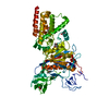
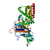
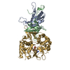
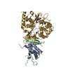
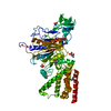
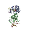
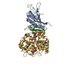
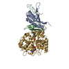
| Title | Structural characterization for the nucleotide binding ability of subunit A with AMP-PNP of the A1AO ATP synthase | ||||||
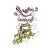
 Components Components | A-TYPE ATP SYNTHASE CATALYTIC SUBUNIT A | ||||||
 Keywords Keywords | HYDROLASE | ||||||
| Function / homology |  Function and homology information Function and homology informationintron homing / intein-mediated protein splicing / proton motive force-driven plasma membrane ATP synthesis / proton-transporting ATPase activity, rotational mechanism / H+-transporting two-sector ATPase / proton-transporting ATP synthase activity, rotational mechanism / endonuclease activity / Hydrolases; Acting on ester bonds / hydrolase activity / ATP binding / plasma membrane Similarity search - Function | ||||||
| Biological species |   Pyrococcus horikoshii (archaea) Pyrococcus horikoshii (archaea) | ||||||
| Method |  X-RAY DIFFRACTION / SYNCHROTRON / MOLECULAR REPLACEMENT / molecular replacement / Resolution: 2.4 Å X-RAY DIFFRACTION / SYNCHROTRON / MOLECULAR REPLACEMENT / molecular replacement / Resolution: 2.4 Å | ||||||
 Authors Authors | Manimekalai, S.M.S. / Kumar, A. / Balakrishna, A.M. / Jeyakanthan, J. / Gruber, G. | ||||||
 Citation Citation | Journal: J.Mol.Biol. / Year: 2010 Title: Nucleotide binding states of subunit A of the A-ATP synthase and the implication of P-loop switch in evolution. Authors: Kumar, A. / Manimekalai, M.S. / Balakrishna, A.M. / Jeyakanthan, J. / Gruber, G. #1: Journal: Acta Crystallogr.,Sect.D / Year: 2006 Title: Structure of the catalytic nucleotide-binding subunit A of A-type ATP synthase from Pyrococcus horikoshii reveals a novel domain related to the peripheral stalk. Authors: Maegawa, Y. / Morita, H. / Iyaguchi, D. / Yao, M. / Watanabe, N. / Tanaka, I. | ||||||
| History |
|
- Structure visualization
| Structure viewer | Molecule: MolmilJmol/JSmol |
|---|
- Downloads & links
-Download
| PDBx/mmCIF format | 3i4l.cif.gz | 129.9 KB | Display | PDBx/mmCIF format |
|---|---|---|---|---|
| PDB format | pdb3i4l.ent.gz | 97.8 KB | Display | PDB format |
| PDBx/mmJSON format | 3i4l.json.gz | Tree view | PDBx/mmJSON format | |
| Others |  Other downloads Other downloads |
-Validation report
| Arichive directory | https://data.pdbj.org/pub/pdb/validation_reports/i4/3i4lftp://data.pdbj.org/pub/pdb/validation_reports/i4/3i4l | HTTPS FTP |
|---|
-Related structure data
| Related structure data |  3i72C  3i73C  3ikjC  1vdzS C: citing same article ( S: Starting model for refinement |
|---|---|
| Similar structure data |
-Links
 PDBj
PDBj


- Assembly
| Deposited unit | 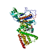
| ||||||||
|---|---|---|---|---|---|---|---|---|---|
| 1 |
| ||||||||
| Unit cell |
| ||||||||
| Details | Biological unit is the same as asymmetric unit |
-Components
-Protein , 1 types, 1 molecules A
| #1: Protein | Mass: 65775.805 Da / Num. of mol.: 1 / Fragment: UNP residues 1-240, 617-964 / Mutation: G79R Source method: isolated from a genetically manipulated source Source: (gene. exp.) Pyrococcus horikoshii (archaea) / Strain: OT3 / Plasmid: pET22b(+)-His6 / Production host:  References: UniProt: O57728, H+-transporting two-sector ATPase |
|---|
-Non-polymers , 5 types, 358 molecules 


| #2: Chemical | ChemComp-ANP /  Mass: 506.196 Da / Num. of mol.: 1 / Source method: obtained synthetically / Formula: C10H17N6O12P3 / Comment: AMP-PNP, energy-carrying molecule analogue*YM Mass: 506.196 Da / Num. of mol.: 1 / Source method: obtained synthetically / Formula: C10H17N6O12P3 / Comment: AMP-PNP, energy-carrying molecule analogue*YM | ||||||
|---|---|---|---|---|---|---|---|
| #3: Chemical |  Mass: 118.174 Da / Num. of mol.: 3 / Source method: obtained synthetically / Formula: C6H14O2 / Comment: precipitant*YM Mass: 118.174 Da / Num. of mol.: 3 / Source method: obtained synthetically / Formula: C6H14O2 / Comment: precipitant*YM#4: Chemical | ChemComp-ACY / |  Mass: 60.052 Da / Num. of mol.: 1 / Source method: obtained synthetically / Formula: C2H4O2 Mass: 60.052 Da / Num. of mol.: 1 / Source method: obtained synthetically / Formula: C2H4O2#5: Chemical | ChemComp-TRS / |  Mass: 122.143 Da / Num. of mol.: 1 / Source method: obtained synthetically / Formula: C4H12NO3 / Comment: pH buffer*YM Mass: 122.143 Da / Num. of mol.: 1 / Source method: obtained synthetically / Formula: C4H12NO3 / Comment: pH buffer*YM#6: Water | ChemComp-HOH / | Mass: 18.015 Da / Num. of mol.: 352 / Source method: isolated from a natural source / Formula: H2O |
-Experimental details
-Experiment
| Experiment | Method: X-RAY DIFFRACTION / Number of used crystals: 1 |
|---|
- Sample preparation
| Crystal | Density Matthews: 3.29 Å3/Da / Density % sol: 62.61 % / Mosaicity: 0.362 ° |
|---|---|
| Crystal grow | Temperature: 291 K / Method: vapor diffusion, hanging drop / pH: 4.5 Details: 50% (v/v) MPD, 0.1M acetate (pH 4.5), vapor diffusion, hanging drop, temperature 291K |
-Data collection
| Diffraction | Mean temperature: 100 K |
|---|---|
| Diffraction source | Source: SYNCHROTRON / Site: SPring-8  / Beamline: BL12B2 / Wavelength: 1 Å / Beamline: BL12B2 / Wavelength: 1 Å |
| Detector | Type: MARMOSAIC 225 mm CCD / Detector: CCD / Date: Oct 20, 2008 / Details: mirrors |
| Radiation | Monochromator: GRAPHITE / Protocol: SINGLE WAVELENGTH / Monochromatic (M) / Laue (L): M / Scattering type: x-ray |
| Radiation wavelength | Wavelength: 1 Å / Relative weight: 1 |
| Reflection | Resolution: 2.4→50 Å / Num. all: 34880 / Num. obs: 34852 / % possible obs: 99.7 % / Observed criterion σ(F): 0 / Observed criterion σ(I): 0 / Redundancy: 14.4 % / Biso Wilson estimate: 65.87 Å2 / Rmerge(I) obs: 0.068 / Χ2: 0.657 / Net I/σ(I): 32.12 |
| Reflection shell | Resolution: 2.4→2.49 Å / Redundancy: 14.1 % / Rmerge(I) obs: 0.459 / Mean I/σ(I) obs: 4.17 / Num. unique all: 3420 / Χ2: 0.491 / % possible all: 100 |
-Phasing
| Phasing | Method: molecular replacement | |||||||||
|---|---|---|---|---|---|---|---|---|---|---|
| Phasing MR | Model details: Phaser MODE: MR_AUTO
|
- Processing
| Software |
| ||||||||||||||||||||||||||||||||||||||||||||||||||||||||||||||||||||||||||||||||||||||||||
|---|---|---|---|---|---|---|---|---|---|---|---|---|---|---|---|---|---|---|---|---|---|---|---|---|---|---|---|---|---|---|---|---|---|---|---|---|---|---|---|---|---|---|---|---|---|---|---|---|---|---|---|---|---|---|---|---|---|---|---|---|---|---|---|---|---|---|---|---|---|---|---|---|---|---|---|---|---|---|---|---|---|---|---|---|---|---|---|---|---|---|---|
| Refinement | Method to determine structure: MOLECULAR REPLACEMENT Starting model: 1vdz Resolution: 2.4→31.14 Å / Cor.coef. Fo:Fc: 0.938 / Cor.coef. Fo:Fc free: 0.931 / WRfactor Rfree: 0.261 / WRfactor Rwork: 0.227 / Occupancy max: 1 / Occupancy min: 0.5 / FOM work R set: 0.813 / SU B: 6.577 / SU ML: 0.155 / SU R Cruickshank DPI: 0.298 / SU Rfree: 0.226 / Cross valid method: THROUGHOUT / σ(F): 0 / ESU R: 0.298 / ESU R Free: 0.226 / Stereochemistry target values: MAXIMUM LIKELIHOOD / Details: HYDROGENS HAVE BEEN ADDED IN THE RIDING POSITIONS
| ||||||||||||||||||||||||||||||||||||||||||||||||||||||||||||||||||||||||||||||||||||||||||
| Solvent computation | Ion probe radii: 0.8 Å / Shrinkage radii: 0.8 Å / VDW probe radii: 1.2 Å / Solvent model: MASK | ||||||||||||||||||||||||||||||||||||||||||||||||||||||||||||||||||||||||||||||||||||||||||
| Displacement parameters | Biso max: 135.29 Å2 / Biso mean: 64.471 Å2 / Biso min: 25.96 Å2
| ||||||||||||||||||||||||||||||||||||||||||||||||||||||||||||||||||||||||||||||||||||||||||
| Refinement step | Cycle: LAST / Resolution: 2.4→31.14 Å
| ||||||||||||||||||||||||||||||||||||||||||||||||||||||||||||||||||||||||||||||||||||||||||
| Refine LS restraints |
| ||||||||||||||||||||||||||||||||||||||||||||||||||||||||||||||||||||||||||||||||||||||||||
| LS refinement shell | Resolution: 2.4→2.462 Å / Total num. of bins used: 20
|
