 Movie
Movie Controller

[English] 日本語
 Yorodumi
- PDB-4gn2: Crystal Structure of OXA-45, a Class D beta-lactamase with extend... -
+ Open data

- Basic information
| Entry | Database: PDB / ID: 4gn2 | ||||||
|---|---|---|---|---|---|---|---|
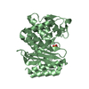
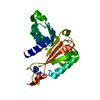
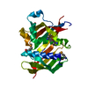
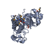
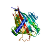
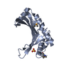
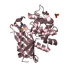
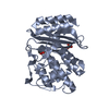
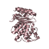
| Title | Crystal Structure of OXA-45, a Class D beta-lactamase with extended spectrum activity | ||||||
 Components Components | Oxacillinase | ||||||
 Keywords Keywords | HYDROLASE / beta-lactamase / antibiotic resistance / lysine carbamylation / alpha/beta | ||||||
| Function / homology |  Function and homology information Function and homology informationpenicillin binding / cell wall organization / beta-lactamase activity / beta-lactamase / plasma membrane Similarity search - Function | ||||||
| Biological species |   Pseudomonas aeruginosa (bacteria) Pseudomonas aeruginosa (bacteria) | ||||||
| Method |  X-RAY DIFFRACTION / MOLECULAR REPLACEMENT / molecular replacement / Resolution: 2.01 Å X-RAY DIFFRACTION / MOLECULAR REPLACEMENT / molecular replacement / Resolution: 2.01 Å | ||||||
 Authors Authors | Martin, J.D. / Xiong, X.L. / Catto, L.E. / Toleman, M.A. / Walsh, T.R. / Clarke, A.R. / Spencer, J. | ||||||
 Citation Citation | Journal: To be Published Title: Structural and Kinetic Characterization of OXA-45, a Class D beta-Lactamase with Extended Spectrum activity Authors: Martin, J.D. / Xiong, X.L. / Catto, L.E. / Toleman, M.A. / Walsh, T.R. / Clarke, A.R. / Spencer, J. | ||||||
| History |
|
- Structure visualization
| Structure viewer | Molecule: MolmilJmol/JSmol |
|---|
- Downloads & links
-Download
| PDBx/mmCIF format | 4gn2.cif.gz | 114.7 KB | Display | PDBx/mmCIF format |
|---|---|---|---|---|
| PDB format | pdb4gn2.ent.gz | 87 KB | Display | PDB format |
| PDBx/mmJSON format | 4gn2.json.gz | Tree view | PDBx/mmJSON format | |
| Others |  Other downloads Other downloads |
-Validation report
| Summary document | 4gn2_validation.pdf.gz | 422.7 KB | Display | wwPDB validaton report |
|---|---|---|---|---|
| Full document | 4gn2_full_validation.pdf.gz | 423.1 KB | Display | |
| Data in XML | 4gn2_validation.xml.gz | 13.7 KB | Display | |
| Data in CIF | 4gn2_validation.cif.gz | 20.4 KB | Display | |
| Arichive directory | https://data.pdbj.org/pub/pdb/validation_reports/gn/4gn2ftp://data.pdbj.org/pub/pdb/validation_reports/gn/4gn2 | HTTPS FTP |
-Related structure data
| Related structure data |  1m6kS S: Starting model for refinement |
|---|---|
| Similar structure data |

-Links
 PDBj
PDBj- Assembly
| Deposited unit | 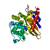
| ||||||||
|---|---|---|---|---|---|---|---|---|---|
| 1 |
| ||||||||
| Unit cell |
|
-Components
| #1: Protein | Mass: 25956.439 Da / Num. of mol.: 1 Source method: isolated from a genetically manipulated source Source: (gene. exp.) Pseudomonas aeruginosa (bacteria) / Strain: 07-406 / Gene: blaoxa45, oxa45 / Plasmid: pK18 / Production host: |
|---|---|
| #2: Water | ChemComp-HOH /  Mass: 18.015 Da / Num. of mol.: 281 / Source method: isolated from a natural source / Formula: H2O Mass: 18.015 Da / Num. of mol.: 281 / Source method: isolated from a natural source / Formula: H2O |
-Experimental details
-Experiment
| Experiment | Method: X-RAY DIFFRACTION / Number of used crystals: 1 |
|---|
- Sample preparation
| Crystal | Density Matthews: 2.25 Å3/Da / Density % sol: 45.33 % |
|---|---|
| Crystal grow | Temperature: 293 K / Method: vapor diffusion, hanging drop / pH: 7.5 Details: 0.1 M HEPES pH 7.5 22% PEG 4K 10% 2-propanol, VAPOR DIFFUSION, HANGING DROP, temperature 293K |
-Data collection
| Diffraction | Mean temperature: 100 K | ||||||||||||||||||||||||||||||||||||||||||||||||||||||||||||||||||
|---|---|---|---|---|---|---|---|---|---|---|---|---|---|---|---|---|---|---|---|---|---|---|---|---|---|---|---|---|---|---|---|---|---|---|---|---|---|---|---|---|---|---|---|---|---|---|---|---|---|---|---|---|---|---|---|---|---|---|---|---|---|---|---|---|---|---|---|
| Diffraction source | Source: ROTATING ANODE / Type: BRUKER AXS MICROSTAR / Wavelength: 1.5418 Å | ||||||||||||||||||||||||||||||||||||||||||||||||||||||||||||||||||
| Detector | Type: BRUKER SMART 6000 / Detector: CCD / Date: Feb 5, 2005 | ||||||||||||||||||||||||||||||||||||||||||||||||||||||||||||||||||
| Radiation | Protocol: SINGLE WAVELENGTH / Monochromatic (M) / Laue (L): M / Scattering type: x-ray | ||||||||||||||||||||||||||||||||||||||||||||||||||||||||||||||||||
| Radiation wavelength | Wavelength: 1.5418 Å / Relative weight: 1 | ||||||||||||||||||||||||||||||||||||||||||||||||||||||||||||||||||
| Reflection | Resolution: 2.01→30 Å / Num. obs: 16275 / % possible obs: 100 % / Rmerge(I) obs: 0.141 / Χ2: 1.026 / Net I/σ(I): 5.4 | ||||||||||||||||||||||||||||||||||||||||||||||||||||||||||||||||||
| Reflection shell |
|
-Phasing
| Phasing | Method: molecular replacement |
|---|
- Processing
| Software |
| ||||||||||||||||||||||||||||||||||||||||||||||||||||||||||||
|---|---|---|---|---|---|---|---|---|---|---|---|---|---|---|---|---|---|---|---|---|---|---|---|---|---|---|---|---|---|---|---|---|---|---|---|---|---|---|---|---|---|---|---|---|---|---|---|---|---|---|---|---|---|---|---|---|---|---|---|---|---|
| Refinement | Method to determine structure: MOLECULAR REPLACEMENT Starting model: 1M6K Resolution: 2.01→27.226 Å / Cor.coef. Fo:Fc: 0.954 / Cor.coef. Fo:Fc free: 0.928 / WRfactor Rfree: 0.1853 / WRfactor Rwork: 0.1461 / Occupancy max: 1 / Occupancy min: 0.5 / FOM work R set: 0.8564 / SU B: 8.579 / SU ML: 0.108 / SU Rfree: 0.1638 / Cross valid method: THROUGHOUT / σ(F): 0 / ESU R Free: 0.164 / Stereochemistry target values: MAXIMUM LIKELIHOOD / Details: U VALUES : REFINED INDIVIDUALLY
| ||||||||||||||||||||||||||||||||||||||||||||||||||||||||||||
| Solvent computation | Ion probe radii: 0.8 Å / Shrinkage radii: 0.8 Å / VDW probe radii: 1.2 Å / Solvent model: MASK | ||||||||||||||||||||||||||||||||||||||||||||||||||||||||||||
| Displacement parameters | Biso max: 72.82 Å2 / Biso mean: 18.1965 Å2 / Biso min: 8.43 Å2
| ||||||||||||||||||||||||||||||||||||||||||||||||||||||||||||
| Refinement step | Cycle: LAST / Resolution: 2.01→27.226 Å
| ||||||||||||||||||||||||||||||||||||||||||||||||||||||||||||
| Refine LS restraints |
| ||||||||||||||||||||||||||||||||||||||||||||||||||||||||||||
| LS refinement shell | Resolution: 2.01→2.062 Å / Total num. of bins used: 20
|
