 Movie
Movie Controller


+ Open data

- Basic information
| Entry | Database: PDB / ID: 1m6k | ||||||
|---|---|---|---|---|---|---|---|
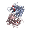
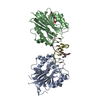
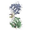
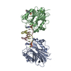
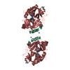
| Title | Structure of the OXA-1 class D beta-lactamase | ||||||
 Components Components | beta-lactamase OXA-1 | ||||||
 Keywords Keywords | HYDROLASE / side chain modification / lysine carbamylation / hydrolysis | ||||||
| Function / homology |  Function and homology information Function and homology informationpenicillin binding / antibiotic catabolic process / beta-lactamase activity / beta-lactamase / periplasmic space / response to antibiotic Similarity search - Function | ||||||
| Biological species |  | ||||||
| Method |  X-RAY DIFFRACTION / SYNCHROTRON / MOLECULAR REPLACEMENT / Resolution: 1.5 Å X-RAY DIFFRACTION / SYNCHROTRON / MOLECULAR REPLACEMENT / Resolution: 1.5 Å | ||||||
 Authors Authors | Sun, T. / Nukaga, M. / Mayama, K. / Braswell, E.H. / Knox, J.R. | ||||||
 Citation Citation | Journal: PROTEIN SCI. / Year: 2003 Title: Comparison of beta-lactamases of classes A and D: 1.5A crystallographic structure of the class D OXA-1 oxacillinase Authors: Sun, T. / Nukaga, M. / Mayama, K. / Braswell, E.H. / Knox, J.R. #1: Journal: Acta Crystallogr.,Sect.D / Year: 2001Title: Crystallization and preliminary X-ray study of OXA-1, a class D beta-lactamase Authors: Sun, T. / Nukaga, M. / Mayama, K. / Crichlow, G.V. / Kuzin, A.P. / Knox, J.R. | ||||||
| History |
| ||||||
| Remark 999 | SEQUENCE AUTHORS STATE THAT THE MATURE, CRYSTALLIZED PROTEIN STARTS AT RESIDUE 18 AS CONFIRMED BY ...SEQUENCE AUTHORS STATE THAT THE MATURE, CRYSTALLIZED PROTEIN STARTS AT RESIDUE 18 AS CONFIRMED BY SEQUENCING. RESIDUES 16 AND 17 ARE IN THE SIGNAL SEQUENCE. AUTHORS ALSO STATE THAT ARG 128 IS AN ERROR IN THE SWISSPROT SEQUENCE AND THAT GLY 128 IS THE CORRECT RESIDUE. |
- Structure visualization
| Structure viewer | Molecule: MolmilJmol/JSmol |
|---|
- Downloads & links
-Download
| PDBx/mmCIF format | 1m6k.cif.gz | 123.2 KB | Display | PDBx/mmCIF format |
|---|---|---|---|---|
| PDB format | pdb1m6k.ent.gz | 95.5 KB | Display | PDB format |
| PDBx/mmJSON format | 1m6k.json.gz | Tree view | PDBx/mmJSON format | |
| Others |  Other downloads Other downloads |
-Validation report
| Arichive directory | https://data.pdbj.org/pub/pdb/validation_reports/m6/1m6kftp://data.pdbj.org/pub/pdb/validation_reports/m6/1m6k | HTTPS FTP |
|---|
-Related structure data


-Links
 PDBj
PDBj- Assembly
| Deposited unit | 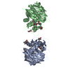
| ||||||||
|---|---|---|---|---|---|---|---|---|---|
| 1 |
| ||||||||
| Unit cell |
|
-Components
| #1: Protein | Mass: 28203.832 Da / Num. of mol.: 2 Source method: isolated from a genetically manipulated source Source: (gene. exp.) #2: Chemical | ChemComp-MPD / (   Mass: 118.174 Da / Num. of mol.: 10 / Source method: obtained synthetically / Formula: C6H14O2 / Comment: precipitant*YM Mass: 118.174 Da / Num. of mol.: 10 / Source method: obtained synthetically / Formula: C6H14O2 / Comment: precipitant*YM#3: Water | ChemComp-HOH / |  Mass: 18.015 Da / Num. of mol.: 559 / Source method: isolated from a natural source / Formula: H2O Mass: 18.015 Da / Num. of mol.: 559 / Source method: isolated from a natural source / Formula: H2OHas protein modification | Y | |
|---|
-Experimental details
-Experiment
| Experiment | Method: X-RAY DIFFRACTION / Number of used crystals: 2 |
|---|
- Sample preparation
| Crystal | Density Matthews: 2.24 Å3/Da / Density % sol: 44 % | ||||||||||||||||||||||||||||||
|---|---|---|---|---|---|---|---|---|---|---|---|---|---|---|---|---|---|---|---|---|---|---|---|---|---|---|---|---|---|---|---|
| Crystal grow | Temperature: 293 K / Method: vapor diffusion, sitting drop / pH: 7.5 Details: PEG 8000 (10/20%), 50 mM HEPES, pH 7.5, VAPOR DIFFUSION, SITTING DROP, temperature 293K | ||||||||||||||||||||||||||||||
| Crystal grow | *PLUS Details: used macroseeding | ||||||||||||||||||||||||||||||
| Components of the solutions | *PLUS
|
-Data collection
| Diffraction |
| ||||||||||||||||||
|---|---|---|---|---|---|---|---|---|---|---|---|---|---|---|---|---|---|---|---|
| Diffraction source |
| ||||||||||||||||||
| Detector |
| ||||||||||||||||||
| Radiation |
| ||||||||||||||||||
| Radiation wavelength |
| ||||||||||||||||||
| Reflection | Resolution: 1.49→50 Å / Num. all: 69837 / Num. obs: 69837 / % possible obs: 86.5 % / Observed criterion σ(F): 0 / Observed criterion σ(I): -3 / Redundancy: 2.6 % / Biso Wilson estimate: 12.8 Å2 / Rmerge(I) obs: 0.049 / Rsym value: 0.049 / Net I/σ(I): 23.6 | ||||||||||||||||||
| Reflection shell | Resolution: 1.5→1.55 Å / Redundancy: 1.5 % / Rmerge(I) obs: 0.123 / Mean I/σ(I) obs: 5.9 / Num. unique all: 2979 / Rsym value: 0.123 / % possible all: 36.9 | ||||||||||||||||||
| Reflection | *PLUS Highest resolution: 1.5 Å / Lowest resolution: 50 Å / Num. obs: 69838 / Redundancy: 2.62 % / Num. measured all: 183270 | ||||||||||||||||||
| Reflection shell | *PLUS % possible obs: 36.9 % / Redundancy: 1.53 % / Num. unique obs: 2979 / Num. measured obs: 4561 / Mean I/σ(I) obs: 5.3 |

- Processing
| Software |
| ||||||||||||||||||||||||||||||||||||
|---|---|---|---|---|---|---|---|---|---|---|---|---|---|---|---|---|---|---|---|---|---|---|---|---|---|---|---|---|---|---|---|---|---|---|---|---|---|
| Refinement | Method to determine structure: MOLECULAR REPLACEMENT Starting model: PDB entries 1FOF and 1E4D Resolution: 1.5→48.21 Å / Rfactor Rfree error: 0.005 / Data cutoff high absF: 500 / Data cutoff high rms absF: 500 / Isotropic thermal model: isotropic B-factors / Cross valid method: THROUGHOUT / σ(F): 0 / σ(I): 0 / Stereochemistry target values: Engh & Huber
| ||||||||||||||||||||||||||||||||||||
| Displacement parameters | Biso mean: 16.4 Å2 | ||||||||||||||||||||||||||||||||||||
| Refine analyze |
| ||||||||||||||||||||||||||||||||||||
| Refinement step | Cycle: LAST / Resolution: 1.5→48.21 Å
| ||||||||||||||||||||||||||||||||||||
| Refine LS restraints |
| ||||||||||||||||||||||||||||||||||||
| LS refinement shell | Resolution: 1.5→1.55 Å / Rfactor Rfree error: 0.022 / Total num. of bins used: 1
| ||||||||||||||||||||||||||||||||||||
| Refinement | *PLUS Highest resolution: 1.5 Å / Lowest resolution: 75 Å / Num. reflection obs: 67388 / Rfactor Rfree: 0.204 / Rfactor Rwork: 0.183 | ||||||||||||||||||||||||||||||||||||
| Solvent computation | *PLUS | ||||||||||||||||||||||||||||||||||||
| Displacement parameters | *PLUS | ||||||||||||||||||||||||||||||||||||
| Refine LS restraints | *PLUS
| ||||||||||||||||||||||||||||||||||||
| LS refinement shell | *PLUS Rfactor Rfree: 0.222 / Rfactor Rwork: 0.215 |
