 Movie
Movie Controller

[English] 日本語
 Yorodumi
- PDB-1ro8: Structural analysis of the sialyltransferase CstII from Campyloba... -
+ Open data

- Basic information
| Entry | Database: PDB / ID: 1ro8 | ||||||
|---|---|---|---|---|---|---|---|
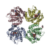
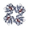
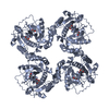
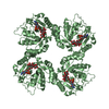
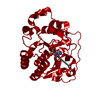
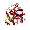
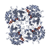
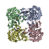
| Title | Structural analysis of the sialyltransferase CstII from Campylobacter jejuni in complex with a substrate analogue, cytidine-5'-monophosphate | ||||||
 Components Components | alpha-2,3/8-sialyltransferase | ||||||
 Keywords Keywords | TRANSFERASE / mixed alpha/beta / Rossmann fold | ||||||
| Function / homology |  Function and homology information Function and homology information | ||||||
| Biological species |   Campylobacter jejuni (Campylobacter) Campylobacter jejuni (Campylobacter) | ||||||
| Method |  X-RAY DIFFRACTION / SYNCHROTRON / MAD / Resolution: 2.05 Å X-RAY DIFFRACTION / SYNCHROTRON / MAD / Resolution: 2.05 Å | ||||||
 Authors Authors | Chiu, C.P. / Watts, A.G. / Lairson, L.L. / Gilbert, M. / Lim, D. / Wakarchuk, W.W. / Withers, S.G. / Strynadka, N.C. | ||||||
 Citation Citation | Journal: Nat.Struct.Mol.Biol. / Year: 2004 Title: Structural analysis of the sialyltransferase CstII from Campylobacter jejuni in complex with a substrate analog. Authors: Chiu, C.P. / Watts, A.G. / Lairson, L.L. / Gilbert, M. / Lim, D. / Wakarchuk, W.W. / Withers, S.G. / Strynadka, N.C. | ||||||
| History |
|
- Structure visualization
| Structure viewer | Molecule: MolmilJmol/JSmol |
|---|
- Downloads & links
-Download
| PDBx/mmCIF format | 1ro8.cif.gz | 111.6 KB | Display | PDBx/mmCIF format |
|---|---|---|---|---|
| PDB format | pdb1ro8.ent.gz | 85.6 KB | Display | PDB format |
| PDBx/mmJSON format | 1ro8.json.gz | Tree view | PDBx/mmJSON format | |
| Others |  Other downloads Other downloads |
-Validation report
| Arichive directory | https://data.pdbj.org/pub/pdb/validation_reports/ro/1ro8ftp://data.pdbj.org/pub/pdb/validation_reports/ro/1ro8 | HTTPS FTP |
|---|
-Related structure data

-Links
 PDBj
PDBj- Assembly
| Deposited unit | 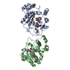
| ||||||||
|---|---|---|---|---|---|---|---|---|---|
| 1 | 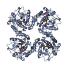
| ||||||||
| 2 | 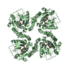
| ||||||||
| Unit cell |
| ||||||||
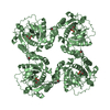
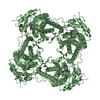
| Details | The biological assembly is a tetramer generated from one molecule in the asymmetric unit by the operations: |
-Components
| #1: Protein | Mass: 30940.596 Da / Num. of mol.: 2 / Mutation: I53S, E222G Source method: isolated from a genetically manipulated source Source: (gene. exp.) Campylobacter jejuni (Campylobacter) / Gene: cst / Plasmid: pET28a / Species (production host): Escherichia coli / Production host: References: UniProt: Q9LAK3, Transferases; Glycosyltransferases; Transferring other glycosyl groups #2: Chemical |   Mass: 323.197 Da / Num. of mol.: 2 / Source method: obtained synthetically / Formula: C9H14N3O8P Mass: 323.197 Da / Num. of mol.: 2 / Source method: obtained synthetically / Formula: C9H14N3O8P#3: Water | ChemComp-HOH / |  Mass: 18.015 Da / Num. of mol.: 64 / Source method: isolated from a natural source / Formula: H2O Mass: 18.015 Da / Num. of mol.: 64 / Source method: isolated from a natural source / Formula: H2OHas protein modification | Y | |
|---|
-Experimental details
-Experiment
| Experiment | Method: X-RAY DIFFRACTION / Number of used crystals: 1 |
|---|
- Sample preparation
| Crystal | Density Matthews: 2.21 Å3/Da / Density % sol: 44.34 % | |||||||||||||||||||||||||||||||||||||||||||||||||
|---|---|---|---|---|---|---|---|---|---|---|---|---|---|---|---|---|---|---|---|---|---|---|---|---|---|---|---|---|---|---|---|---|---|---|---|---|---|---|---|---|---|---|---|---|---|---|---|---|---|---|
| Crystal grow | Temperature: 291 K / Method: vapor diffusion, hanging drop / pH: 7.5 Details: PEG 6000, MPD, Tris, pH 7.5, VAPOR DIFFUSION, HANGING DROP, temperature 291K | |||||||||||||||||||||||||||||||||||||||||||||||||
| Crystal grow | *PLUS Method: vapor diffusion, hanging drop | |||||||||||||||||||||||||||||||||||||||||||||||||
| Components of the solutions | *PLUS
|
-Data collection
| Diffraction | Mean temperature: 100 K | ||||||||||||
|---|---|---|---|---|---|---|---|---|---|---|---|---|---|
| Diffraction source | Source: SYNCHROTRON / Site: NSLS  / Beamline: X25 / Wavelength: 0.97912, 0.97961, 0.97900 / Beamline: X25 / Wavelength: 0.97912, 0.97961, 0.97900 | ||||||||||||
| Detector | Type: ADSC QUANTUM 315 / Detector: CCD / Date: Sep 25, 2002 / Details: mirrors | ||||||||||||
| Radiation | Monochromator: Si(111), (220) and W-B4C multilayer / Protocol: MAD / Monochromatic (M) / Laue (L): M / Scattering type: x-ray | ||||||||||||
| Radiation wavelength |
| ||||||||||||
| Reflection | Resolution: 2→30 Å / Num. obs: 62730 / % possible obs: 95 % / Observed criterion σ(F): 2 / Observed criterion σ(I): 2 / Redundancy: 5.8 % / Biso Wilson estimate: 11.9 Å2 / Rsym value: 0.057 / Net I/σ(I): 23.9 | ||||||||||||
| Reflection shell | Resolution: 2→2.18 Å / Mean I/σ(I) obs: 10.6 / Rsym value: 0.091 / % possible all: 95 |
- Processing
| Software |
| |||||||||||||||||||||||||
|---|---|---|---|---|---|---|---|---|---|---|---|---|---|---|---|---|---|---|---|---|---|---|---|---|---|---|
| Refinement | Method to determine structure: MAD / Resolution: 2.05→28.95 Å / Rfactor Rfree error: 0.005 / Data cutoff high absF: 2324601.26 / Data cutoff low absF: 0 / Isotropic thermal model: RESTRAINED / Cross valid method: THROUGHOUT / σ(F): 0 / Stereochemistry target values: Engh & Huber
| |||||||||||||||||||||||||
| Solvent computation | Solvent model: FLAT MODEL / Bsol: 42.5553 Å2 / ksol: 0.356015 e/Å3 | |||||||||||||||||||||||||
| Displacement parameters | Biso mean: 40.9 Å2
| |||||||||||||||||||||||||
| Refine analyze |
| |||||||||||||||||||||||||
| Refinement step | Cycle: LAST / Resolution: 2.05→28.95 Å
| |||||||||||||||||||||||||
| Refine LS restraints |
| |||||||||||||||||||||||||
| LS refinement shell | Resolution: 2.05→2.18 Å / Rfactor Rfree error: 0.015 / Total num. of bins used: 6
| |||||||||||||||||||||||||
| Xplor file |
| |||||||||||||||||||||||||
| Refinement | *PLUS Highest resolution: 2.1 Å / Lowest resolution: 30 Å / % reflection Rfree: 5 % / Rfactor Rfree: 0.256 / Rfactor Rwork: 0.219 | |||||||||||||||||||||||||
| Solvent computation | *PLUS | |||||||||||||||||||||||||
| Displacement parameters | *PLUS | |||||||||||||||||||||||||
| Refine LS restraints | *PLUS
|
