 Movie
Movie Controller

[English] 日本語
 Yorodumi
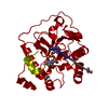
- PDB-2x61: Crystal structure of the sialyltransferase CST-II in complex with... -
+ Open data

- Basic information
| Entry | Database: PDB / ID: 2x61 | |||||||||
|---|---|---|---|---|---|---|---|---|---|---|
| Title | Crystal structure of the sialyltransferase CST-II in complex with trisaccharide acceptor and CMP | |||||||||
 Components Components | ALPHA-2,3-/2,8-SIALYLTRANSFERASE | |||||||||
 Keywords Keywords | TRANSFERASE / GTA / GLYCOSYLTRANSFERASE | |||||||||
| Function / homology |  Function and homology information Function and homology information | |||||||||
| Biological species |   CAMPYLOBACTER JEJUNI (Campylobacter) CAMPYLOBACTER JEJUNI (Campylobacter) | |||||||||
| Method |  X-RAY DIFFRACTION / SYNCHROTRON / MOLECULAR REPLACEMENT / Resolution: 1.95 Å X-RAY DIFFRACTION / SYNCHROTRON / MOLECULAR REPLACEMENT / Resolution: 1.95 Å | |||||||||
 Authors Authors | Lee, H.J. / Lairson, L.L. / Rich, J.R. / Wakarchuk, W.W. / Withers, S.G. / Strynadka, N.C.J. | |||||||||
 Citation Citation | Journal: J.Biol.Chem. / Year: 2011 Title: Structural and Kinetic Analysis of Substrate Binding to the Sialyltransferase Cst-II from Campylobacter Jejuni. Authors: Lee, H.J. / Lairson, L.L. / Rich, J.R. / Lameignere, E. / Wakarchuk, W.W. / Withers, S.G. / Strynadka, N.C. | |||||||||
| History |
|
- Structure visualization
| Structure viewer | Molecule: MolmilJmol/JSmol |
|---|
- Downloads & links
-Download
| PDBx/mmCIF format | 2x61.cif.gz | 223.6 KB | Display | PDBx/mmCIF format |
|---|---|---|---|---|
| PDB format | pdb2x61.ent.gz | 179.4 KB | Display | PDB format |
| PDBx/mmJSON format | 2x61.json.gz | Tree view | PDBx/mmJSON format | |
| Others |  Other downloads Other downloads |
-Validation report
| Arichive directory | https://data.pdbj.org/pub/pdb/validation_reports/x6/2x61ftp://data.pdbj.org/pub/pdb/validation_reports/x6/2x61 | HTTPS FTP |
|---|
-Related structure data
| Related structure data |  2x62C 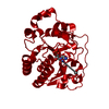 2x63C 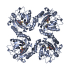 1ro8S S: Starting model for refinement C: citing same article ( |
|---|---|
| Similar structure data |


-Links
 PDBj
PDBj- Assembly
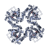
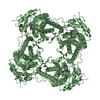
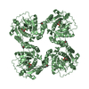
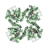
| Deposited unit | 
| ||||||||
|---|---|---|---|---|---|---|---|---|---|
| 1 | 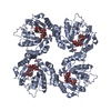
| ||||||||
| 2 | 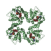
| ||||||||
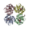
| Unit cell |
|
-Components
-Protein / Sugars , 2 types, 4 molecules AB
| #1: Protein | Mass: 30403.531 Da / Num. of mol.: 2 / Fragment: RESIDUES 1-258 / Mutation: YES Source method: isolated from a genetically manipulated source Source: (gene. exp.) CAMPYLOBACTER JEJUNI (Campylobacter) / Strain: OH4384 / Production host: References: UniProt: Q9LAK3, Transferases; Glycosyltransferases; Transferring other glycosyl groups #2: Polysaccharide | Source method: isolated from a genetically manipulated source |
|---|
-Non-polymers , 5 types, 271 molecules 

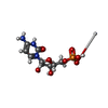


| #3: Chemical |  Mass: 59.044 Da / Num. of mol.: 3 / Source method: obtained synthetically / Formula: C2H3O2 Mass: 59.044 Da / Num. of mol.: 3 / Source method: obtained synthetically / Formula: C2H3O2#4: Chemical | ChemComp-MPD / ( |  Mass: 118.174 Da / Num. of mol.: 1 / Source method: obtained synthetically / Formula: C6H14O2 / Comment: precipitant*YM Mass: 118.174 Da / Num. of mol.: 1 / Source method: obtained synthetically / Formula: C6H14O2 / Comment: precipitant*YM#5: Chemical |  Type: RNA linking / Mass: 324.204 Da / Num. of mol.: 2 / Source method: obtained synthetically / Formula: C9H15N3O8P Type: RNA linking / Mass: 324.204 Da / Num. of mol.: 2 / Source method: obtained synthetically / Formula: C9H15N3O8P#6: Chemical |  Mass: 62.068 Da / Num. of mol.: 2 / Source method: obtained synthetically / Formula: C2H6O2 Mass: 62.068 Da / Num. of mol.: 2 / Source method: obtained synthetically / Formula: C2H6O2#7: Water | ChemComp-HOH / | Mass: 18.015 Da / Num. of mol.: 263 / Source method: isolated from a natural source / Formula: H2O |
|---|
-Details
| Compound details | ENGINEERED RESIDUE IN CHAIN A, ILE 53 TO SER ENGINEERED RESIDUE IN CHAIN A, GLU 222 TO GLY ...ENGINEERED |
|---|
-Experimental details
-Experiment
| Experiment | Method: X-RAY DIFFRACTION / Number of used crystals: 1 |
|---|
- Sample preparation
| Crystal | Density Matthews: 2.52 Å3/Da / Density % sol: 51 % / Description: NONE |
|---|---|
| Crystal grow | pH: 7.5 Details: 100 MM HEPES5, PH 7.5, 8% (W/V) POLYETHYLENE GLYCOL 6000, AND 5% (V/V) 2-METHYL-2, 4-PENTANEDIOL |
-Data collection
| Diffraction | Mean temperature: 100 K |
|---|---|
| Diffraction source | Source: SYNCHROTRON / Site: CLSI  / Beamline: 08ID-1 / Wavelength: 0.979 / Beamline: 08ID-1 / Wavelength: 0.979 |
| Detector | Type: MARRESEARCH / Detector: CCD / Date: Dec 10, 2008 / Details: VERTICALLY FOCUSSING MIRROR |
| Radiation | Monochromator: DOUBLE CRYSTAL / Protocol: SINGLE WAVELENGTH / Monochromatic (M) / Laue (L): M / Scattering type: x-ray |
| Radiation wavelength | Wavelength: 0.979 Å / Relative weight: 1 |
| Reflection | Resolution: 1.95→40 Å / Num. obs: 46782 / % possible obs: 99.6 % / Observed criterion σ(I): 5 / Redundancy: 6.3 % / Rmerge(I) obs: 0.08 / Net I/σ(I): 38.3 |
| Reflection shell | Resolution: 1.95→2.02 Å / Redundancy: 5.6 % / Rmerge(I) obs: 0.37 / Mean I/σ(I) obs: 5 / % possible all: 96.9 |
- Processing
| Software |
| ||||||||||||||||||||||||||||||||||||||||||||||||||||||||||||||||||||||||||||||||||||||||||||||||||||||||||||||||||||||||||||||||||||||||||||||||||||||||||||||||||||||||||||||||||||||
|---|---|---|---|---|---|---|---|---|---|---|---|---|---|---|---|---|---|---|---|---|---|---|---|---|---|---|---|---|---|---|---|---|---|---|---|---|---|---|---|---|---|---|---|---|---|---|---|---|---|---|---|---|---|---|---|---|---|---|---|---|---|---|---|---|---|---|---|---|---|---|---|---|---|---|---|---|---|---|---|---|---|---|---|---|---|---|---|---|---|---|---|---|---|---|---|---|---|---|---|---|---|---|---|---|---|---|---|---|---|---|---|---|---|---|---|---|---|---|---|---|---|---|---|---|---|---|---|---|---|---|---|---|---|---|---|---|---|---|---|---|---|---|---|---|---|---|---|---|---|---|---|---|---|---|---|---|---|---|---|---|---|---|---|---|---|---|---|---|---|---|---|---|---|---|---|---|---|---|---|---|---|---|---|
| Refinement | Method to determine structure: MOLECULAR REPLACEMENT Starting model: PDB ENTRY 1RO8 Resolution: 1.95→40 Å / Cor.coef. Fo:Fc: 0.967 / Cor.coef. Fo:Fc free: 0.951 / SU B: 6.39 / SU ML: 0.085 / Cross valid method: THROUGHOUT / ESU R: 0.135 / ESU R Free: 0.125 / Stereochemistry target values: MAXIMUM LIKELIHOOD / Details: HYDROGENS HAVE BEEN ADDED IN THE RIDING POSITIONS
| ||||||||||||||||||||||||||||||||||||||||||||||||||||||||||||||||||||||||||||||||||||||||||||||||||||||||||||||||||||||||||||||||||||||||||||||||||||||||||||||||||||||||||||||||||||||
| Solvent computation | Ion probe radii: 0.8 Å / Shrinkage radii: 0.8 Å / VDW probe radii: 1.4 Å / Solvent model: MASK | ||||||||||||||||||||||||||||||||||||||||||||||||||||||||||||||||||||||||||||||||||||||||||||||||||||||||||||||||||||||||||||||||||||||||||||||||||||||||||||||||||||||||||||||||||||||
| Displacement parameters | Biso mean: 36.496 Å2
| ||||||||||||||||||||||||||||||||||||||||||||||||||||||||||||||||||||||||||||||||||||||||||||||||||||||||||||||||||||||||||||||||||||||||||||||||||||||||||||||||||||||||||||||||||||||
| Refinement step | Cycle: LAST / Resolution: 1.95→40 Å
| ||||||||||||||||||||||||||||||||||||||||||||||||||||||||||||||||||||||||||||||||||||||||||||||||||||||||||||||||||||||||||||||||||||||||||||||||||||||||||||||||||||||||||||||||||||||
| Refine LS restraints |
|