 Movie
Movie Controller

[English] 日本語
 Yorodumi
- PDB-2drj: Xray structure of alpha-2,3/8-sialyltransferase CstII F91Y mutant -
+ Open data

- Basic information
| Entry | Database: PDB / ID: 2drj | ||||||
|---|---|---|---|---|---|---|---|
| Title | Xray structure of alpha-2,3/8-sialyltransferase CstII F91Y mutant | ||||||
 Components Components | Alpha-2,3/8-sialyltransferase | ||||||
 Keywords Keywords | TRANSFERASE / mixed alpha/beta | ||||||
| Function / homology |  Function and homology information Function and homology information | ||||||
| Biological species |   Campylobacter jejuni (Campylobacter) Campylobacter jejuni (Campylobacter) | ||||||
| Method |  X-RAY DIFFRACTION / MOLECULAR REPLACEMENT / Resolution: 2.25 Å X-RAY DIFFRACTION / MOLECULAR REPLACEMENT / Resolution: 2.25 Å | ||||||
 Authors Authors | Chiu, C.P.C. / Strynadka, N.C.J. | ||||||
 Citation Citation | Journal: Nat.Methods / Year: 2006 Title: High-throughput screening methodology for the directed evolution of glycosyltransferases Authors: Aharoni, A. / Thieme, K. / Chiu, C.P.C. / Buchini, S. / Lairson, L.L. / Chen, H. / Strynadka, N.C.J. / Wakarchuk, W.W. / Withers, S.G. | ||||||
| History |
|
- Structure visualization
| Structure viewer | Molecule: MolmilJmol/JSmol |
|---|
- Downloads & links
-Download
| PDBx/mmCIF format | 2drj.cif.gz | 66.6 KB | Display | PDBx/mmCIF format |
|---|---|---|---|---|
| PDB format | pdb2drj.ent.gz | 47.8 KB | Display | PDB format |
| PDBx/mmJSON format | 2drj.json.gz | Tree view | PDBx/mmJSON format | |
| Others |  Other downloads Other downloads |
-Validation report
| Arichive directory | https://data.pdbj.org/pub/pdb/validation_reports/dr/2drjftp://data.pdbj.org/pub/pdb/validation_reports/dr/2drj | HTTPS FTP |
|---|
-Related structure data
| Related structure data | 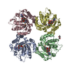 1ro7S S: Starting model for refinement |
|---|---|
| Similar structure data |


-Links
 PDBj
PDBj
- Assembly
| Deposited unit | 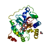
| ||||||||
|---|---|---|---|---|---|---|---|---|---|
| 1 | 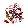
| ||||||||
| Unit cell |
| ||||||||
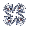
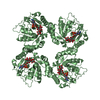
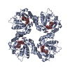
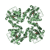
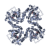
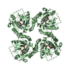
| Details | The biological assembly is a tetramer generated from the molecule in the asymmetric unit by the operations: -x,-y,z; -y,x,z; y,-x,z. |
-Components
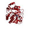
| #1: Protein | Mass: 30815.912 Da / Num. of mol.: 1 / Fragment: sialyltransferase residues 1-259 / Mutation: F91Y Source method: isolated from a genetically manipulated source Source: (gene. exp.) Campylobacter jejuni (Campylobacter) / Plasmid: pET / Species (production host): Escherichia coli / Production host: References: UniProt: Q9LAK3, Transferases; Glycosyltransferases; Transferring other glycosyl groups | ||||
|---|---|---|---|---|---|
| #2: Chemical | ChemComp-CSF / 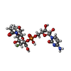  Type: RNA linking / Mass: 632.442 Da / Num. of mol.: 1 / Source method: obtained synthetically / Formula: C20H30FN4O16P Type: RNA linking / Mass: 632.442 Da / Num. of mol.: 1 / Source method: obtained synthetically / Formula: C20H30FN4O16P | ||||
| #3: Chemical |   Mass: 60.095 Da / Num. of mol.: 2 / Source method: obtained synthetically / Formula: C3H8O Mass: 60.095 Da / Num. of mol.: 2 / Source method: obtained synthetically / Formula: C3H8O#4: Water | ChemComp-HOH / |  Mass: 18.015 Da / Num. of mol.: 92 / Source method: isolated from a natural source / Formula: H2O Mass: 18.015 Da / Num. of mol.: 92 / Source method: isolated from a natural source / Formula: H2OHas protein modification | N | |
-Experimental details
-Experiment
| Experiment | Method: X-RAY DIFFRACTION / Number of used crystals: 1 |
|---|
- Sample preparation
| Crystal | Density Matthews: 2.46 Å3/Da / Density % sol: 49.9 % |
|---|---|
| Crystal grow | Temperature: 283 K / Method: vapor diffusion, hanging drop / pH: 7.5 Details: 0.1M TEA pH 7.5, 20% PEG 400, 10% isopropanol, VAPOR DIFFUSION, HANGING DROP, temperature 283K |
-Data collection
| Diffraction | Mean temperature: 100 K |
|---|---|
| Diffraction source | Source: ROTATING ANODE / Type: RIGAKU / Wavelength: 1.514 Å |
| Detector | Type: MAR scanner 345 mm plate / Detector: IMAGE PLATE / Date: Nov 23, 2005 / Details: mirrors |
| Radiation | Monochromator: Ni filter / Protocol: SINGLE WAVELENGTH / Monochromatic (M) / Laue (L): M / Scattering type: x-ray |
| Radiation wavelength | Wavelength: 1.514 Å / Relative weight: 1 |
| Reflection | Resolution: 2.25→25 Å / Num. obs: 13458 / % possible obs: 94.3 % / Observed criterion σ(F): 2 / Observed criterion σ(I): 2 / Redundancy: 3.9 % / Rmerge(I) obs: 0.066 / Χ2: 0.986 / Net I/σ(I): 23.1 |
| Reflection shell | Resolution: 2.25→2.33 Å / Redundancy: 3.1 % / Rmerge(I) obs: 0.185 / Num. unique all: 926 / Χ2: 0.948 / % possible all: 65.4 |
- Processing
| Software |
| ||||||||||||||||||||||||||||||||||||||||||||||||||||||||||||||||||||||||||||||||||||||||||
|---|---|---|---|---|---|---|---|---|---|---|---|---|---|---|---|---|---|---|---|---|---|---|---|---|---|---|---|---|---|---|---|---|---|---|---|---|---|---|---|---|---|---|---|---|---|---|---|---|---|---|---|---|---|---|---|---|---|---|---|---|---|---|---|---|---|---|---|---|---|---|---|---|---|---|---|---|---|---|---|---|---|---|---|---|---|---|---|---|---|---|---|
| Refinement | Method to determine structure: MOLECULAR REPLACEMENT Starting model: PDB entry 1RO7 Resolution: 2.25→23.81 Å / Cor.coef. Fo:Fc: 0.945 / Cor.coef. Fo:Fc free: 0.895 / SU B: 5.545 / SU ML: 0.144 / Cross valid method: THROUGHOUT / σ(F): 0 / ESU R: 0.305 / ESU R Free: 0.242 / Stereochemistry target values: MAXIMUM LIKELIHOOD
| ||||||||||||||||||||||||||||||||||||||||||||||||||||||||||||||||||||||||||||||||||||||||||
| Solvent computation | Ion probe radii: 0.8 Å / Shrinkage radii: 0.8 Å / VDW probe radii: 1.4 Å / Solvent model: MASK | ||||||||||||||||||||||||||||||||||||||||||||||||||||||||||||||||||||||||||||||||||||||||||
| Displacement parameters | Biso mean: 33.254 Å2
| ||||||||||||||||||||||||||||||||||||||||||||||||||||||||||||||||||||||||||||||||||||||||||
| Refinement step | Cycle: LAST / Resolution: 2.25→23.81 Å
| ||||||||||||||||||||||||||||||||||||||||||||||||||||||||||||||||||||||||||||||||||||||||||
| Refine LS restraints |
| ||||||||||||||||||||||||||||||||||||||||||||||||||||||||||||||||||||||||||||||||||||||||||
| LS refinement shell | Resolution: 2.251→2.309 Å / Total num. of bins used: 20
|
