 Movie
Movie Controller


+ Open data

- Basic information
| Entry | Database: PDB / ID: 4ggv | ||||||
|---|---|---|---|---|---|---|---|
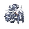
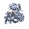
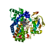
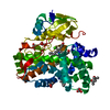
| Title | Crystal Structure of HmtT Involved in Himastatin Biosynthesis | ||||||
 Components Components | Cytochrome P450 superfamily protein | ||||||
 Keywords Keywords | OXIDOREDUCTASE / Cysteine-ligand loop / Hydrolase | ||||||
| Function / homology |  Function and homology information Function and homology informationcholest-4-en-3-one 26-monooxygenase activity / steroid hydroxylase activity / cholesterol catabolic process / iron ion binding / heme binding Similarity search - Function | ||||||
| Biological species |  Streptomyces himastatinicus (bacteria) Streptomyces himastatinicus (bacteria) | ||||||
| Method |  X-RAY DIFFRACTION / SYNCHROTRON / MOLECULAR REPLACEMENT / Resolution: 2.331 Å X-RAY DIFFRACTION / SYNCHROTRON / MOLECULAR REPLACEMENT / Resolution: 2.331 Å | ||||||
 Authors Authors | Zhang, H. / Chen, J. / Wang, H. / Zhang, H. | ||||||
 Citation Citation | Journal: Febs Lett. / Year: 2013 Title: Structural analysis of HmtT and HmtN involved in the tailoring steps of himastatin biosynthesis Authors: Zhang, H. / Chen, J. / Wang, H. / Xie, Y. / Ju, J. / Yan, Y. / Zhang, H. | ||||||
| History |
|
- Structure visualization
| Structure viewer | Molecule: MolmilJmol/JSmol |
|---|
- Downloads & links
-Download
| PDBx/mmCIF format | 4ggv.cif.gz | 94.7 KB | Display | PDBx/mmCIF format |
|---|---|---|---|---|
| PDB format | pdb4ggv.ent.gz | 70.8 KB | Display | PDB format |
| PDBx/mmJSON format | 4ggv.json.gz | Tree view | PDBx/mmJSON format | |
| Others |  Other downloads Other downloads |
-Validation report
| Arichive directory | https://data.pdbj.org/pub/pdb/validation_reports/gg/4ggvftp://data.pdbj.org/pub/pdb/validation_reports/gg/4ggv | HTTPS FTP |
|---|
-Related structure data
| Related structure data | 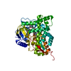 2jjoS S: Starting model for refinement |
|---|---|
| Similar structure data |


-Links
 PDBj
PDBj
- Assembly
| Deposited unit | 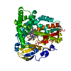
| ||||||||
|---|---|---|---|---|---|---|---|---|---|
| 1 |
| ||||||||
| Unit cell |
| ||||||||
| Components on special symmetry positions |
|
-Components
| #1: Protein | Mass: 46477.496 Da / Num. of mol.: 1 Source method: isolated from a genetically manipulated source Source: (gene. exp.) Streptomyces himastatinicus (bacteria) / Strain: ATCC 53653 / Gene: hmtT, SSOG_07642 / Plasmid: pET28a / Production host: References: UniProt: D9WMR2, Oxidoreductases; Acting on paired donors, with incorporation or reduction of molecular oxygen; With NADH or NADPH as one donor, and incorporation of one atom of oxygen into the other donor |
|---|---|
| #2: Chemical | ChemComp-HEM /   Mass: 616.487 Da / Num. of mol.: 1 / Source method: obtained synthetically / Formula: C34H32FeN4O4 Mass: 616.487 Da / Num. of mol.: 1 / Source method: obtained synthetically / Formula: C34H32FeN4O4 |
| #3: Water | ChemComp-HOH /  Mass: 18.015 Da / Num. of mol.: 150 / Source method: isolated from a natural source / Formula: H2O Mass: 18.015 Da / Num. of mol.: 150 / Source method: isolated from a natural source / Formula: H2O |
-Experimental details
-Experiment
| Experiment | Method: X-RAY DIFFRACTION / Number of used crystals: 1 |
|---|
- Sample preparation
| Crystal | Density Matthews: 2.3 Å3/Da / Density % sol: 46.45 % |
|---|---|
| Crystal grow | Temperature: 277.15 K / Method: vapor diffusion, sitting drop / pH: 4.6 Details: 8%(w/v) PEG4000, 3%(v/v) Dimethyl Sulfoxide, 0.1M Sodium acetate trihydrate, pH 4.6, VAPOR DIFFUSION, SITTING DROP, temperature 277.15K |
-Data collection
| Diffraction | Mean temperature: 77.15 K |
|---|---|
| Diffraction source | Source: SYNCHROTRON / Site: SSRF  / Beamline: BL17U / Wavelength: 0.979228 Å / Beamline: BL17U / Wavelength: 0.979228 Å |
| Detector | Type: ADSC QUANTUM 315 / Detector: CCD / Date: May 2, 2012 |
| Radiation | Protocol: SINGLE WAVELENGTH / Monochromatic (M) / Laue (L): M / Scattering type: x-ray |
| Radiation wavelength | Wavelength: 0.979228 Å / Relative weight: 1 |
| Reflection | Resolution: 2.331→44.1244 Å / Num. all: 18771 / Num. obs: 17864 / % possible obs: 99.9578 % / Observed criterion σ(F): 3 / Observed criterion σ(I): 2.4 / Redundancy: 4.3 % / Biso Wilson estimate: 24.89 Å2 / Rmerge(I) obs: 0.071 |
| Reflection shell | Resolution: 2.331→2.37 Å / % possible all: 99.9 |
- Processing
| Software |
| ||||||||||||||||||||||||||||||||||||||||||||||||
|---|---|---|---|---|---|---|---|---|---|---|---|---|---|---|---|---|---|---|---|---|---|---|---|---|---|---|---|---|---|---|---|---|---|---|---|---|---|---|---|---|---|---|---|---|---|---|---|---|---|
| Refinement | Method to determine structure: MOLECULAR REPLACEMENT Starting model: 2JJO Resolution: 2.331→44.124 Å / SU ML: 0.24 / σ(F): 1.36 / Phase error: 25.14 / Stereochemistry target values: ML
| ||||||||||||||||||||||||||||||||||||||||||||||||
| Solvent computation | Shrinkage radii: 0.6 Å / VDW probe radii: 0.9 Å / Solvent model: FLAT BULK SOLVENT MODEL / Bsol: 27.525 Å2 / ksol: 0.337 e/Å3 | ||||||||||||||||||||||||||||||||||||||||||||||||
| Displacement parameters |
| ||||||||||||||||||||||||||||||||||||||||||||||||
| Refinement step | Cycle: LAST / Resolution: 2.331→44.124 Å
| ||||||||||||||||||||||||||||||||||||||||||||||||
| Refine LS restraints |
| ||||||||||||||||||||||||||||||||||||||||||||||||
| LS refinement shell | Refine-ID: X-RAY DIFFRACTION / Total num. of bins used: 7
|
