 Movie
Movie Controller

[English] 日本語
 Yorodumi
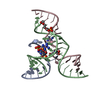
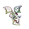
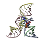
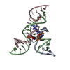
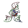
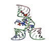
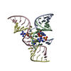
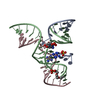
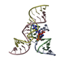
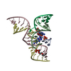
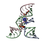
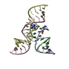
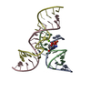
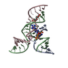
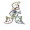
- PDB-4g6s: Minimal Hairpin Ribozyme in the Transition State with A38P Variation -
+ Open data

- Basic information
| Entry | Database: PDB / ID: 4g6s | ||||||
|---|---|---|---|---|---|---|---|
| Title | Minimal Hairpin Ribozyme in the Transition State with A38P Variation | ||||||
 Components Components |
| ||||||
 Keywords Keywords | RNA / Structure-activity relationship / Nucleic Acid Conformation | ||||||
| Function / homology | COBALT HEXAMMINE(III) / RNA / RNA (> 10) Function and homology information Function and homology information | ||||||
| Method |  X-RAY DIFFRACTION / SYNCHROTRON / FOURIER SYNTHESIS / Resolution: 2.84 Å X-RAY DIFFRACTION / SYNCHROTRON / FOURIER SYNTHESIS / Resolution: 2.84 Å | ||||||
 Authors Authors | Liberman, J.A. / Jenkins, J.L. / Krucinska, J. / Wedekind, J.E. | ||||||
 Citation Citation | Journal: J.Am.Chem.Soc. / Year: 2012 Title: A Transition-State Interaction Shifts Nucleobase Ionization toward Neutrality To Facilitate Small Ribozyme Catalysis. Authors: Liberman, J.A. / Guo, M. / Jenkins, J.L. / Krucinska, J. / Chen, Y. / Carey, P.R. / Wedekind, J.E. | ||||||
| History |
|
- Structure visualization
| Structure viewer | Molecule: MolmilJmol/JSmol |
|---|
- Downloads & links
-Download
| PDBx/mmCIF format | 4g6s.cif.gz | 45.9 KB | Display | PDBx/mmCIF format |
|---|---|---|---|---|
| PDB format | pdb4g6s.ent.gz | 32.4 KB | Display | PDB format |
| PDBx/mmJSON format | 4g6s.json.gz | Tree view | PDBx/mmJSON format | |
| Others |  Other downloads Other downloads |
-Validation report
| Arichive directory | https://data.pdbj.org/pub/pdb/validation_reports/g6/4g6sftp://data.pdbj.org/pub/pdb/validation_reports/g6/4g6s | HTTPS FTP |
|---|
-Related structure data


-Links
 PDBj
PDBj


- Assembly
| Deposited unit | 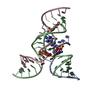
| ||||||||
|---|---|---|---|---|---|---|---|---|---|
| 1 |
| ||||||||
| Unit cell |
| ||||||||
| Components on special symmetry positions |
|
-Components
-RNA chain , 3 types, 3 molecules ABC
| #1: RNA chain | Mass: 4036.471 Da / Num. of mol.: 1 / Source method: obtained synthetically |
|---|---|
| #2: RNA chain | Mass: 9762.989 Da / Num. of mol.: 1 / Source method: obtained synthetically |
| #3: RNA chain | Mass: 5976.554 Da / Num. of mol.: 1 / Source method: obtained synthetically |
-Non-polymers , 3 types, 6 molecules 


| #4: Chemical | ChemComp-SO4 /  Mass: 96.063 Da / Num. of mol.: 1 / Source method: obtained synthetically / Formula: SO4 Mass: 96.063 Da / Num. of mol.: 1 / Source method: obtained synthetically / Formula: SO4 | ||
|---|---|---|---|
| #5: Chemical |  Mass: 161.116 Da / Num. of mol.: 2 / Source method: obtained synthetically / Formula: CoH18N6 Mass: 161.116 Da / Num. of mol.: 2 / Source method: obtained synthetically / Formula: CoH18N6#6: Water | ChemComp-HOH / | Mass: 18.015 Da / Num. of mol.: 3 / Source method: isolated from a natural source / Formula: H2O |
-Experimental details
-Experiment
| Experiment | Method: X-RAY DIFFRACTION / Number of used crystals: 1 |
|---|
- Sample preparation
| Crystal | Density Matthews: 4.32 Å3/Da / Density % sol: 71.55 % |
|---|---|
| Crystal grow | Temperature: 295 K / Method: vapor diffusion, hanging drop / pH: 7 Details: 22% (w/v) PEG 2000 MME, 0.1M HEPES pH=7.0, 0.25 M Lithium Sulfate, 1mM Cobalt Hexamine, 2mM spermadine, vapor diffusion, Hanging Drop, temperature 295K |
-Data collection
| Diffraction | Mean temperature: 100 K | |||||||||||||||||||||||||||||||||||||||||||||||||||||||||||||||||||||||||||||
|---|---|---|---|---|---|---|---|---|---|---|---|---|---|---|---|---|---|---|---|---|---|---|---|---|---|---|---|---|---|---|---|---|---|---|---|---|---|---|---|---|---|---|---|---|---|---|---|---|---|---|---|---|---|---|---|---|---|---|---|---|---|---|---|---|---|---|---|---|---|---|---|---|---|---|---|---|---|---|
| Diffraction source | Source: SYNCHROTRON / Site: SSRL  / Beamline: BL11-1 / Wavelength: 0.97885 Å / Beamline: BL11-1 / Wavelength: 0.97885 Å | |||||||||||||||||||||||||||||||||||||||||||||||||||||||||||||||||||||||||||||
| Detector | Type: MARMOSAIC 325 mm CCD / Detector: CCD / Date: Feb 5, 2010 / Details: Rh coated flat mirror | |||||||||||||||||||||||||||||||||||||||||||||||||||||||||||||||||||||||||||||
| Radiation | Monochromator: Si(111) / Protocol: SINGLE WAVELENGTH / Scattering type: x-ray | |||||||||||||||||||||||||||||||||||||||||||||||||||||||||||||||||||||||||||||
| Radiation wavelength | Wavelength: 0.97885 Å / Relative weight: 1 | |||||||||||||||||||||||||||||||||||||||||||||||||||||||||||||||||||||||||||||
| Reflection | Resolution: 2.84→50 Å / Num. obs: 8742 / % possible obs: 98.7 % / Redundancy: 6.9 % / Rmerge(I) obs: 0.052 / Χ2: 1.36 / Net I/σ(I): 16 | |||||||||||||||||||||||||||||||||||||||||||||||||||||||||||||||||||||||||||||
| Reflection shell |
|
- Processing
| Software |
| ||||||||||||||||||||||||||||
|---|---|---|---|---|---|---|---|---|---|---|---|---|---|---|---|---|---|---|---|---|---|---|---|---|---|---|---|---|---|
| Refinement | Method to determine structure: FOURIER SYNTHESIS / Resolution: 2.84→30.097 Å / Occupancy max: 1 / Occupancy min: 0.49 / SU ML: 0.33 / σ(F): 0 / Phase error: 29.88 / Stereochemistry target values: ML
| ||||||||||||||||||||||||||||
| Solvent computation | Shrinkage radii: 0.83 Å / VDW probe radii: 1.1 Å / Solvent model: FLAT BULK SOLVENT MODEL / Bsol: 32.875 Å2 / ksol: 0.244 e/Å3 | ||||||||||||||||||||||||||||
| Displacement parameters | Biso max: 138.16 Å2 / Biso mean: 89.0083 Å2 / Biso min: 61.87 Å2
| ||||||||||||||||||||||||||||
| Refinement step | Cycle: LAST / Resolution: 2.84→30.097 Å
| ||||||||||||||||||||||||||||
| Refine LS restraints |
| ||||||||||||||||||||||||||||
| LS refinement shell | Refine-ID: X-RAY DIFFRACTION / Total num. of bins used: 3
|
