 Movie
Movie Controller

[English] 日本語
 Yorodumi
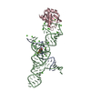
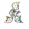
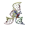
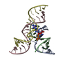
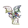
- PDB-2p7e: Vanadate at the Active Site of a Small Ribozyme Suggests a Role f... -
+ Open data

- Basic information
| Entry | Database: PDB / ID: 2p7e | ||||||
|---|---|---|---|---|---|---|---|
| Title | Vanadate at the Active Site of a Small Ribozyme Suggests a Role for Water in Transition-State Stabilization | ||||||
 Components Components |
| ||||||
 Keywords Keywords | RNA / hairpin ribozyme / vanadate / reaction intermediate / transition-state stabilization / active site waters / induced fit | ||||||
| Function / homology | COBALT HEXAMMINE(III) / DNA/RNA hybrid / DNA/RNA hybrid (> 10) / RNA / RNA (> 10) Function and homology information Function and homology information | ||||||
| Method |  X-RAY DIFFRACTION / SYNCHROTRON / FOURIER SYNTHESIS / Resolution: 2.05 Å X-RAY DIFFRACTION / SYNCHROTRON / FOURIER SYNTHESIS / Resolution: 2.05 Å | ||||||
 Authors Authors | Torelli, A.T. / Krucinska, J. / Wedekind, J.E. | ||||||
 Citation Citation | Journal: Rna / Year: 2007 Title: A comparison of vanadate to a 2'-5' linkage at the active site of a small ribozyme suggests a role for water in transition-state stabilization Authors: Torelli, A.T. / Krucinska, J. / Wedekind, J.E. #1: Journal: Biochemistry / Year: 2006Title: Water in the active site of an all-RNA hairpin ribozyme and effects of Gua8 base variants on the geometry of phosphoryl transfer. Authors: Salter, J. / Krucinska, J. / Alam, S. / Grum-Tokars, V. / Wedekind, J.E. #2: Journal: Biochemistry / Year: 2005Title: Conformational heterogeneity at position U37 of an all-RNA hairpin ribozyme with implications for metal binding and the catalytic structure of the S-turn. Authors: Alam, S. / Grum-Tokars, V. / Krucinska, J. / Kundracik, M.L. / Wedekind, J.E. | ||||||
| History |
|
- Structure visualization
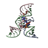
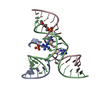
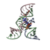
| Structure viewer | Molecule: MolmilJmol/JSmol |
|---|
- Downloads & links
-Download
| PDBx/mmCIF format | 2p7e.cif.gz | 49.5 KB | Display | PDBx/mmCIF format |
|---|---|---|---|---|
| PDB format | pdb2p7e.ent.gz | 34.8 KB | Display | PDB format |
| PDBx/mmJSON format | 2p7e.json.gz | Tree view | PDBx/mmJSON format | |
| Others |  Other downloads Other downloads |
-Validation report
| Arichive directory | https://data.pdbj.org/pub/pdb/validation_reports/p7/2p7eftp://data.pdbj.org/pub/pdb/validation_reports/p7/2p7e | HTTPS FTP |
|---|
-Related structure data
| Related structure data |  2p7dC  2p7fC  1zfr C: citing same article ( S: Starting model for refinement |
|---|---|
| Similar structure data |


-Links
 PDBj
PDBj


- Assembly
| Deposited unit | 
| ||||||||
|---|---|---|---|---|---|---|---|---|---|
| 1 |
| ||||||||
| Unit cell |
| ||||||||
| Components on special symmetry positions |
| ||||||||
| Details | The ribozyme-substrate complex in the asymmetric unit also represents the biological unit |
-Components
-RNA chain , 2 types, 2 molecules AP
| #1: RNA chain | Mass: 1587.893 Da / Num. of mol.: 1 / Source method: obtained synthetically Details: sequence occurs naturally in tobacco ringspot virus satellite RNA |
|---|---|
| #5: RNA chain | Mass: 2501.553 Da / Num. of mol.: 1 / Source method: obtained synthetically Details: sequence occurs naturally in tobacco ringspot virus satellite RNA |
-Ribozyme strand ... , 3 types, 3 molecules BCD
| #2: DNA/RNA hybrid | Mass: 4182.585 Da / Num. of mol.: 1 / Source method: obtained synthetically Details: sequence occurs naturally in tobacco ringspot virus satellite RNA |
|---|---|
| #3: RNA chain | Mass: 5535.445 Da / Num. of mol.: 1 / Source method: obtained synthetically Details: sequence occurs naturally in tobacco ringspot virus satellite RNA |
| #4: RNA chain | Mass: 5991.568 Da / Num. of mol.: 1 / Source method: obtained synthetically Details: sequence occurs naturally in tobacco ringspot virus satellite RNA |
-Non-polymers , 3 types, 64 molecules 


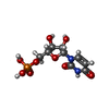
| #6: Chemical | ChemComp-SO4 /  Mass: 96.063 Da / Num. of mol.: 1 / Source method: obtained synthetically / Formula: SO4 Mass: 96.063 Da / Num. of mol.: 1 / Source method: obtained synthetically / Formula: SO4 | ||
|---|---|---|---|
| #7: Chemical |  Mass: 161.116 Da / Num. of mol.: 2 / Source method: obtained synthetically / Formula: CoH18N6 Mass: 161.116 Da / Num. of mol.: 2 / Source method: obtained synthetically / Formula: CoH18N6#8: Water | ChemComp-HOH / | Mass: 18.015 Da / Num. of mol.: 61 / Source method: isolated from a natural source / Formula: H2O |
-Experimental details
-Experiment
| Experiment | Method: X-RAY DIFFRACTION / Number of used crystals: 1 |
|---|
- Sample preparation
| Crystal | Density Matthews: 4.27 Å3/Da / Density % sol: 80.57 % | ||||||||||||||||||||||||||||||||||||||||
|---|---|---|---|---|---|---|---|---|---|---|---|---|---|---|---|---|---|---|---|---|---|---|---|---|---|---|---|---|---|---|---|---|---|---|---|---|---|---|---|---|---|
| Crystal grow | Temperature: 293 K / Method: vapor diffusion, hanging drop / pH: 6 Details: 250 mM Li2SO4, 2.5 mM [Co(NH3)6]Cl3, 2 mM spermidine-HCl, 4.5 mM ammonium metavanadate, 100 mM sodium cacodylate buffer and 20% PEG 2000 MME, pH 6.0, VAPOR DIFFUSION, HANGING DROP, temperature 293K | ||||||||||||||||||||||||||||||||||||||||
| Components of the solutions |
|
-Data collection
| Diffraction | Mean temperature: 100 K |
|---|---|
| Diffraction source | Source: SYNCHROTRON / Site: SSRL  / Beamline: BL9-1 / Wavelength: 0.98789 Å / Beamline: BL9-1 / Wavelength: 0.98789 Å |
| Detector | Type: ADSC QUANTUM 315 / Detector: CCD / Date: Apr 12, 2006 Details: Vertical focusing mirror; single crystal Si(311) bent monochromator (horizontal focusing) |
| Radiation | Monochromator: Side-scattering cuberoot I-beam bent single crystal; asymmetric cut 12.2 degs Protocol: SINGLE WAVELENGTH / Monochromatic (M) / Laue (L): M / Scattering type: x-ray |
| Radiation wavelength | Wavelength: 0.98789 Å / Relative weight: 1 |
| Reflection | Resolution: 2.05→30.67 Å / Num. all: 21610 / Num. obs: 21610 / % possible obs: 96.3 % / Observed criterion σ(I): -3 / Redundancy: 5.45 % / Biso Wilson estimate: 52.6 Å2 / Rsym value: 0.046 / Net I/σ(I): 14.9 |
| Reflection shell | Resolution: 2.05→2.12 Å / Redundancy: 4.86 % / Mean I/σ(I) obs: 3.5 / Rsym value: 0.429 / % possible all: 85.8 |
- Processing
| Software |
| |||||||||||||||||||||||||||
|---|---|---|---|---|---|---|---|---|---|---|---|---|---|---|---|---|---|---|---|---|---|---|---|---|---|---|---|---|
| Refinement | Method to determine structure: FOURIER SYNTHESIS Starting model: PDB ENTRY 1ZFR 1zfr Resolution: 2.05→29.93 Å / Rfactor Rfree error: 0.008 / Data cutoff high absF: 2624271.15 / Data cutoff low absF: 0 / Isotropic thermal model: RESTRAINED / Cross valid method: THROUGHOUT / σ(F): 0 Stereochemistry target values: Parkinson et al. Acta Cryst.D,52,57 (1996)
| |||||||||||||||||||||||||||
| Solvent computation | Solvent model: FLAT MODEL / Bsol: 55.4615 Å2 / ksol: 0.316013 e/Å3 | |||||||||||||||||||||||||||
| Displacement parameters | Biso mean: 61 Å2
| |||||||||||||||||||||||||||
| Refine analyze |
| |||||||||||||||||||||||||||
| Refinement step | Cycle: LAST / Resolution: 2.05→29.93 Å
| |||||||||||||||||||||||||||
| Refine LS restraints |
| |||||||||||||||||||||||||||
| LS refinement shell | Resolution: 2.05→2.18 Å / Rfactor Rfree error: 0.047 / Total num. of bins used: 6
| |||||||||||||||||||||||||||
| Xplor file |
|
