 Movie
Movie Controller

[English] 日本語
 Yorodumi
- PDB-3b5f: Crystal Structure of a Minimally Hinged Hairpin Ribozyme Incorpor... -
+ Open data

- Basic information
| Entry | Database: PDB / ID: 3b5f | ||||||
|---|---|---|---|---|---|---|---|
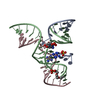
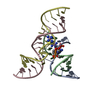
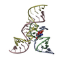
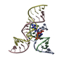
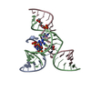
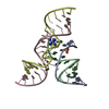
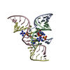
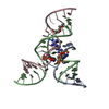
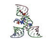
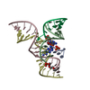
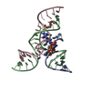
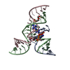
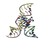
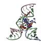
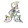
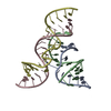
| Title | Crystal Structure of a Minimally Hinged Hairpin Ribozyme Incorporating the Ade38Dap Mutation and a 2',5' Phosphodiester Linkage at the Active Site | ||||||
 Components Components |
| ||||||
 Keywords Keywords | RNA / hairpin ribozyme / 2 / 6 diaminopurine / 2' / 5' phosphodiester linkage | ||||||
| Function / homology | COBALT HEXAMMINE(III) / RNA / RNA (> 10) Function and homology information Function and homology information | ||||||
| Method |  X-RAY DIFFRACTION / SYNCHROTRON / FOURIER SYNTHESIS / Resolution: 2.7 Å X-RAY DIFFRACTION / SYNCHROTRON / FOURIER SYNTHESIS / Resolution: 2.7 Å | ||||||
 Authors Authors | MacElrevey, C. / Krucinska, J. / Wedekind, J.E. | ||||||
 Citation Citation | Journal: Rna / Year: 2008 Title: Structural effects of nucleobase variations at key active site residue Ade38 in the hairpin ribozyme. Authors: MacElrevey, C. / Salter, J.D. / Krucinska, J. / Wedekind, J.E. | ||||||
| History |
|
- Structure visualization
| Structure viewer | Molecule: MolmilJmol/JSmol |
|---|
- Downloads & links
-Download
| PDBx/mmCIF format | 3b5f.cif.gz | 45.8 KB | Display | PDBx/mmCIF format |
|---|---|---|---|---|
| PDB format | pdb3b5f.ent.gz | 32.3 KB | Display | PDB format |
| PDBx/mmJSON format | 3b5f.json.gz | Tree view | PDBx/mmJSON format | |
| Others |  Other downloads Other downloads |
-Validation report
| Arichive directory | https://data.pdbj.org/pub/pdb/validation_reports/b5/3b5fftp://data.pdbj.org/pub/pdb/validation_reports/b5/3b5f | HTTPS FTP |
|---|
-Related structure data
| Related structure data |  3b58C  3b5aC  3b5sC  3b91C  3bbiC  3bbkC  3bbmC 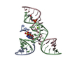 3cr1C  2p7fS S: Starting model for refinement C: citing same article ( |
|---|---|
| Similar structure data |


-Links
 PDBj
PDBj


- Assembly
| Deposited unit | 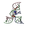
| ||||||||
|---|---|---|---|---|---|---|---|---|---|
| 1 |
| ||||||||
| Unit cell |
|
-Components
| #1: RNA chain | Mass: 4052.470 Da / Num. of mol.: 1 / Source method: obtained synthetically / Details: solid-phase synthesis at Keck Institute, Yale / References: PDB-3B58 | ||
|---|---|---|---|
| #2: RNA chain | Mass: 9762.989 Da / Num. of mol.: 1 / Source method: obtained synthetically Details: solid phase chemical synthesis at Dharmacon, Colorado References: PDB-3B58 | ||
| #3: RNA chain | Mass: 6006.583 Da / Num. of mol.: 1 / Mutation: A38(N6G), U39C / Source method: obtained synthetically Details: solid phase chemical synthesis at Dharmacon, Colorado References: PDB-3B58 | ||
| #4: Chemical |   Mass: 161.116 Da / Num. of mol.: 2 / Source method: obtained synthetically / Formula: CoH18N6 Mass: 161.116 Da / Num. of mol.: 2 / Source method: obtained synthetically / Formula: CoH18N6#5: Water | ChemComp-HOH / |  Mass: 18.015 Da / Num. of mol.: 3 / Source method: isolated from a natural source / Formula: H2O Mass: 18.015 Da / Num. of mol.: 3 / Source method: isolated from a natural source / Formula: H2O |
-Experimental details
-Experiment
| Experiment | Method: X-RAY DIFFRACTION / Number of used crystals: 1 |
|---|
- Sample preparation
| Crystal | Density Matthews: 4.34 Å3/Da / Density % sol: 78 % | ||||||||||||||||||||||||||||||||||||
|---|---|---|---|---|---|---|---|---|---|---|---|---|---|---|---|---|---|---|---|---|---|---|---|---|---|---|---|---|---|---|---|---|---|---|---|---|---|
| Crystal grow | Temperature: 293 K / Method: vapor diffusion, hanging drop / pH: 6.8 Details: PEG 2K MME, lithium sulfate, sodium cacodylate, spermidine, cobalt hexaammine, pH 6.8, VAPOR DIFFUSION, HANGING DROP, temperature 293K | ||||||||||||||||||||||||||||||||||||
| Components of the solutions |
|
-Data collection
| Diffraction | Mean temperature: 100 K |
|---|---|
| Diffraction source | Source: SYNCHROTRON / Site: CHESS  / Beamline: F1 / Wavelength: 0.918 Å / Beamline: F1 / Wavelength: 0.918 Å |
| Detector | Type: ADSC QUANTUM 270 / Detector: CCD / Date: May 10, 2007 Details: BENT TRIANGULAR ASYMMETRIC CUT SI(111) MONOCHROMATOR (PROVIDES HORIZONTAL FOCUSSING), RH-COATED SI MIRROR FOR VERTICAL FOCUSSING |
| Radiation | Monochromator: horizontal bent Si(111) asymmetrically cut w/ water cooled Cu block Protocol: SINGLE WAVELENGTH / Monochromatic (M) / Laue (L): M / Scattering type: x-ray |
| Radiation wavelength | Wavelength: 0.918 Å / Relative weight: 1 |
| Reflection | Resolution: 2.7→40.48 Å / Num. all: 10109 / Num. obs: 9960 / % possible obs: 99.4 % / Redundancy: 7.8 % / Biso Wilson estimate: 79.6 Å2 / Rsym value: 0.05 / Net I/σ(I): 23.8 |
| Reflection shell | Resolution: 2.7→2.8 Å / Redundancy: 6.1 % / Mean I/σ(I) obs: 3 / Num. unique all: 962 / Rsym value: 0.459 / % possible all: 99.9 |
- Processing
| Software |
| |||||||||||||||||||||||||
|---|---|---|---|---|---|---|---|---|---|---|---|---|---|---|---|---|---|---|---|---|---|---|---|---|---|---|
| Refinement | Method to determine structure: FOURIER SYNTHESIS Starting model: 2P7F Resolution: 2.7→38.32 Å / Rfactor Rfree error: 0.008 / Data cutoff high absF: 599805.33 / Data cutoff low absF: 0 / Isotropic thermal model: RESTRAINED / Cross valid method: THROUGHOUT / σ(F): -3 Details: Alternate conformation at U1 of chain A. S9L and N6G are covalently attached to ribozyme
| |||||||||||||||||||||||||
| Solvent computation | Solvent model: FLAT MODEL / Bsol: 60 Å2 / ksol: 0.32 e/Å3 | |||||||||||||||||||||||||
| Displacement parameters | Biso mean: 81.2 Å2
| |||||||||||||||||||||||||
| Refine analyze |
| |||||||||||||||||||||||||
| Refinement step | Cycle: LAST / Resolution: 2.7→38.32 Å
| |||||||||||||||||||||||||
| Refine LS restraints |
| |||||||||||||||||||||||||
| LS refinement shell | Resolution: 2.7→2.97 Å / Rfactor Rfree error: 0.039 / Total num. of bins used: 4
| |||||||||||||||||||||||||
| Xplor file |
|
