 Movie
Movie Controller

[English] 日本語
 Yorodumi
- PDB-2oue: Crystal structure of a junctionless all-RNA hairpin ribozyme at 2... -
+ Open data

- Basic information
| Entry | Database: PDB / ID: 2oue | |||||||||
|---|---|---|---|---|---|---|---|---|---|---|
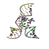
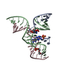
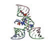
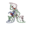
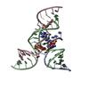
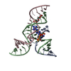
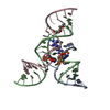
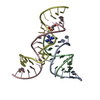
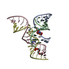
| Title | Crystal structure of a junctionless all-RNA hairpin ribozyme at 2.05 angstroms resolution | |||||||||
 Components Components |
| |||||||||
 Keywords Keywords | RNA / hairpin ribozyme / all-RNA / mutation / low salt / S-turn / E-loop / ribose zipper / catalytic RNA | |||||||||
| Function / homology | COBALT HEXAMMINE(III) / RNA / RNA (> 10) Function and homology information Function and homology information | |||||||||
| Method |  X-RAY DIFFRACTION / SYNCHROTRON / FOURIER SYNTHESIS / Resolution: 2.05 Å X-RAY DIFFRACTION / SYNCHROTRON / FOURIER SYNTHESIS / Resolution: 2.05 Å | |||||||||
 Authors Authors | Wedekind, J.E. | |||||||||
 Citation Citation | Journal: Biochemistry / Year: 2006 Title: Water in the Active Site of an All-RNA Hairpin Ribozyme and Effects of Gua8 Base Variants on the Geometry of Phosphoryl Transfer. Authors: Salter, J. / Krucinska, J. / Alam, S. / Grum-Tokars, V. / Wedekind, J.E. | |||||||||
| History |
|
- Structure visualization
| Structure viewer | Molecule: MolmilJmol/JSmol |
|---|
- Downloads & links
-Download
| PDBx/mmCIF format | 2oue.cif.gz | 47.2 KB | Display | PDBx/mmCIF format |
|---|---|---|---|---|
| PDB format | pdb2oue.ent.gz | 33.8 KB | Display | PDB format |
| PDBx/mmJSON format | 2oue.json.gz | Tree view | PDBx/mmJSON format | |
| Others |  Other downloads Other downloads |
-Validation report
| Arichive directory | https://data.pdbj.org/pub/pdb/validation_reports/ou/2oueftp://data.pdbj.org/pub/pdb/validation_reports/ou/2oue | HTTPS FTP |
|---|
-Related structure data
| Related structure data |  1zftC  1zfvC  1zfxC  2bcyC  2bczC  2fgpC C: citing same article ( |
|---|---|
| Similar structure data |


-Links
 PDBj
PDBj


- Assembly
| Deposited unit | 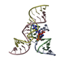
| ||||||||
|---|---|---|---|---|---|---|---|---|---|
| 1 |
| ||||||||
| Unit cell |
|
-Components
-RNA chain , 4 types, 4 molecules ABCD
| #1: RNA chain | Mass: 4066.497 Da / Num. of mol.: 1 / Source method: obtained synthetically Details: Chemically synthesized based on satellite tobacco ringspot virus |
|---|---|
| #2: RNA chain | Mass: 3970.448 Da / Num. of mol.: 1 / Source method: obtained synthetically Details: Chemically synthesized based on satellite tobacco ringspot virus |
| #3: RNA chain | Mass: 5535.445 Da / Num. of mol.: 1 / Source method: obtained synthetically Details: Chemically synthesized based on satellite tobacco ringspot virus |
| #4: RNA chain | Mass: 5991.568 Da / Num. of mol.: 1 / Source method: obtained synthetically Details: Chemically synthesized based on satellite tobacco ringspot virus |
-Non-polymers , 3 types, 87 molecules 


| #5: Chemical | ChemComp-SO4 /  Mass: 96.063 Da / Num. of mol.: 1 / Source method: obtained synthetically / Formula: SO4 Mass: 96.063 Da / Num. of mol.: 1 / Source method: obtained synthetically / Formula: SO4 | ||
|---|---|---|---|
| #6: Chemical |  Mass: 161.116 Da / Num. of mol.: 2 / Source method: obtained synthetically / Formula: CoH18N6 Mass: 161.116 Da / Num. of mol.: 2 / Source method: obtained synthetically / Formula: CoH18N6#7: Water | ChemComp-HOH / | Mass: 18.015 Da / Num. of mol.: 84 / Source method: isolated from a natural source / Formula: H2O |
-Experimental details
-Experiment
| Experiment | Method: X-RAY DIFFRACTION / Number of used crystals: 1 |
|---|
- Sample preparation
| Crystal | Density Matthews: 4.21 Å3/Da / Density % sol: 70.8 % | ||||||||||||||||||||||||||||||||||||||||||||
|---|---|---|---|---|---|---|---|---|---|---|---|---|---|---|---|---|---|---|---|---|---|---|---|---|---|---|---|---|---|---|---|---|---|---|---|---|---|---|---|---|---|---|---|---|---|
| Crystal grow | Temperature: 293 K / Method: vapor diffusion, hanging drop / pH: 6 Details: PEG 2000 MME, LITHIUM SULFATE, cacodylate, spermidine, cobalt hexaamine, pH 6.0, VAPOR DIFFUSION, HANGING DROP, temperature 293K | ||||||||||||||||||||||||||||||||||||||||||||
| Components of the solutions |
|
-Data collection
| Diffraction | Mean temperature: 100 K |
|---|---|
| Diffraction source | Source: SYNCHROTRON / Site: CHESS  / Beamline: A1 / Wavelength: 0.9764 Å / Beamline: A1 / Wavelength: 0.9764 Å |
| Detector | Type: ADSC QUANTUM 210 / Detector: CCD / Date: Nov 28, 2004 Details: BENT TRIANGULAR ASYMMETRIC CUT SI(111) MONOCHROMATOR (PROVIDES HORIZONTAL FOCUSSING), RH-COATED SI MIRROR FOR VERTICAL FOCUSSING |
| Radiation | Monochromator: BENT TRIANGULAR ASYMMETRIC CUT SI(111) MONOCHROMATOR Protocol: SINGLE WAVELENGTH / Monochromatic (M) / Laue (L): M / Scattering type: x-ray |
| Radiation wavelength | Wavelength: 0.9764 Å / Relative weight: 1 |
| Reflection | Resolution: 2.05→30.4 Å / Num. obs: 21699 / % possible obs: 99.3 % / Observed criterion σ(F): 0 / Observed criterion σ(I): -5 / Redundancy: 8.2 % / Biso Wilson estimate: 61.8 Å2 / Net I/σ(I): 24 |
| Reflection shell | Resolution: 2.05→2.12 Å / Redundancy: 4.7 % / Mean I/σ(I) obs: 3.3 / Rsym value: 0.432 / % possible all: 99.1 |
- Processing
| Software |
| |||||||||||||||||||||||||
|---|---|---|---|---|---|---|---|---|---|---|---|---|---|---|---|---|---|---|---|---|---|---|---|---|---|---|
| Refinement | Method to determine structure: FOURIER SYNTHESIS / Resolution: 2.05→30.4 Å / Rfactor Rfree error: 0.006 / Data cutoff high absF: 1422181.38 / Data cutoff high rms absF: 1422181.38 / Data cutoff low absF: 0 / Isotropic thermal model: RESTRAINED / Cross valid method: THROUGHOUT / σ(F): 0 / σ(I): -5 / Stereochemistry target values: Engh & Huber
| |||||||||||||||||||||||||
| Solvent computation | Solvent model: FLAT MODEL / Bsol: 63.093 Å2 / ksol: 0.343432 e/Å3 | |||||||||||||||||||||||||
| Displacement parameters | Biso mean: 69.8 Å2
| |||||||||||||||||||||||||
| Refine analyze |
| |||||||||||||||||||||||||
| Refinement step | Cycle: LAST / Resolution: 2.05→30.4 Å
| |||||||||||||||||||||||||
| Refine LS restraints |
| |||||||||||||||||||||||||
| LS refinement shell | Resolution: 2.05→2.26 Å / Rfactor Rfree error: 0.03 / Total num. of bins used: 4
| |||||||||||||||||||||||||
| Xplor file |
|
