 Movie
Movie Controller

[English] 日本語
 Yorodumi
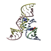
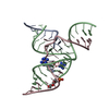
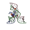
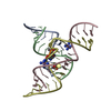
- PDB-2fgp: Crystal structure of a minimal, all RNA hairpin ribozyme with mod... -
+ Open data

- Basic information
| Entry | Database: PDB / ID: 2fgp | ||||||
|---|---|---|---|---|---|---|---|
| Title | Crystal structure of a minimal, all RNA hairpin ribozyme with modifications (g8dap, u39c) at ph 8.6 | ||||||
 Components Components |
| ||||||
 Keywords Keywords | RNA / RIBOZYME / G8 / DIAMINOPURINE / IN-LINE GEOMETRY / MUTANT | ||||||
| Function / homology | COBALT HEXAMMINE(III) / RNA / RNA (> 10) Function and homology information Function and homology information | ||||||
| Method |  X-RAY DIFFRACTION / SYNCHROTRON / FOURIER SYNTHESIS / Resolution: 2.4 Å X-RAY DIFFRACTION / SYNCHROTRON / FOURIER SYNTHESIS / Resolution: 2.4 Å | ||||||
 Authors Authors | Salter, J.D. / Wedekind, J.E. | ||||||
 Citation Citation | Journal: Biochemistry / Year: 2006 Title: Water in the Active Site of an All-RNA Hairpin Ribozyme and Effects of Gua8 Base Variants on the Geometry of Phosphoryl Transfer. Authors: Salter, J.D. / Krucinska, J. / Alam, S. / Grum-Tokars, V. / Wedekind, J.E. #1: Journal: Biochemistry / Year: 2005Title: Conformational Heterogeneity at Position U37 of an All-RNA Hairpin Ribozyme with Implications for Metal Binding and the Catalytic Structure of the S-Turn Authors: Alam, S. / Grum-Tokars, V. / Krucinska, J. / Kundracik, M.L. / Wedekind, J.E. #2: Journal: Acta Crystallogr.,Sect.D / Year: 2003Title: Crystallization and x-ray diffraction analysis of an all-rna u39c mutant of the minimal hairpin ribozyme Authors: Grum-Tokars, V. / Milovanovic, M. / Wedekind, J.E. | ||||||
| History |
|
- Structure visualization
| Structure viewer | Molecule: MolmilJmol/JSmol |
|---|
- Downloads & links
-Download
| PDBx/mmCIF format | 2fgp.cif.gz | 45.9 KB | Display | PDBx/mmCIF format |
|---|---|---|---|---|
| PDB format | pdb2fgp.ent.gz | 32.2 KB | Display | PDB format |
| PDBx/mmJSON format | 2fgp.json.gz | Tree view | PDBx/mmJSON format | |
| Others |  Other downloads Other downloads |
-Validation report
| Arichive directory | https://data.pdbj.org/pub/pdb/validation_reports/fg/2fgpftp://data.pdbj.org/pub/pdb/validation_reports/fg/2fgp | HTTPS FTP |
|---|
-Related structure data
| Related structure data |  1zftC 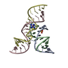 1zfvC  1zfxC  2bcyC  2bczC  2oueC  1zfr C: citing same article ( S: Starting model for refinement |
|---|---|
| Similar structure data |


-Links
 PDBj
PDBj


- Assembly
| Deposited unit | 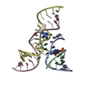
| ||||||||
|---|---|---|---|---|---|---|---|---|---|
| 1 |
| ||||||||
| Unit cell |
|
-Components
-RNA chain , 4 types, 4 molecules ABCD
| #1: RNA chain | Mass: 4052.470 Da / Num. of mol.: 1 / Source method: obtained synthetically / Details: DERIVED FROM SATELLITE TOBACCO RINGSPOT VIRUS |
|---|---|
| #2: RNA chain | Mass: 3969.463 Da / Num. of mol.: 1 / Mutation: g8(N6G) / Source method: obtained synthetically / Details: DERIVED FROM SATELLITE TOBACCO RINGSPOT VIRUS |
| #3: RNA chain | Mass: 5535.445 Da / Num. of mol.: 1 / Source method: obtained synthetically / Details: DERIVED FROM SATELLITE TOBACCO RINGSPOT VIRUS |
| #4: RNA chain | Mass: 5991.568 Da / Num. of mol.: 1 / Mutation: u39c / Source method: obtained synthetically / Details: DERIVED FROM SATELLITE TOBACCO RINGSPOT VIRUS |
-Non-polymers , 2 types, 8 molecules 

| #5: Chemical | ChemComp-NCO /  Mass: 161.116 Da / Num. of mol.: 1 / Source method: obtained synthetically / Formula: CoH18N6 Mass: 161.116 Da / Num. of mol.: 1 / Source method: obtained synthetically / Formula: CoH18N6 |
|---|---|
| #6: Water | ChemComp-HOH / Mass: 18.015 Da / Num. of mol.: 7 / Source method: isolated from a natural source / Formula: H2O |
-Experimental details
-Experiment
| Experiment | Method: X-RAY DIFFRACTION / Number of used crystals: 1 |
|---|
- Sample preparation
| Crystal | Density Matthews: 4.14 Å3/Da / Density % sol: 70.31 % | ||||||||||||||||||||||||||||||||||||||||||||
|---|---|---|---|---|---|---|---|---|---|---|---|---|---|---|---|---|---|---|---|---|---|---|---|---|---|---|---|---|---|---|---|---|---|---|---|---|---|---|---|---|---|---|---|---|---|
| Crystal grow | Temperature: 293 K / Method: vapor diffusion, hanging drop / pH: 8.6 Details: PEG2000 MME, LITHIUM SULFATE, SPERMIDINE, COBALT HEXAMMINE, pH 8.60, VAPOR DIFFUSION, HANGING DROP, temperature 293K | ||||||||||||||||||||||||||||||||||||||||||||
| Components of the solutions |
|
-Data collection
| Diffraction | Mean temperature: 83 K |
|---|---|
| Diffraction source | Source: SYNCHROTRON / Site: CHESS  / Beamline: A1 / Wavelength: 0.9764 / Beamline: A1 / Wavelength: 0.9764 |
| Detector | Type: ADSC QUANTUM 210 / Detector: CCD / Date: May 5, 2005 |
| Radiation | Monochromator: BENT TRIANGULAR ASYMMETRIC CUT SI(111) MONOCHROMATOR, RH- COATED SI FOR VERTICAL FOCUSSING Protocol: SINGLE WAVELENGTH / Monochromatic (M) / Laue (L): M / Scattering type: x-ray |
| Radiation wavelength | Wavelength: 0.9764 Å / Relative weight: 1 |
| Reflection | Resolution: 2.4→44.01 Å / Num. obs: 12161 / % possible obs: 89.8 % / Observed criterion σ(F): 0 / Observed criterion σ(I): -3 / Redundancy: 10.56 % / Biso Wilson estimate: 88.1 Å2 / Rmerge(I) obs: 0.034 / Net I/σ(I): 34.8 |
| Reflection shell | Resolution: 2.4→2.49 Å / Redundancy: 9.25 % / Rmerge(I) obs: 0.458 / Mean I/σ(I) obs: 5.1 / % possible all: 92.8 |
- Processing
| Software |
| ||||||||||||||||||||||||||||||||||||||||||||||||||||||||||||
|---|---|---|---|---|---|---|---|---|---|---|---|---|---|---|---|---|---|---|---|---|---|---|---|---|---|---|---|---|---|---|---|---|---|---|---|---|---|---|---|---|---|---|---|---|---|---|---|---|---|---|---|---|---|---|---|---|---|---|---|---|---|
| Refinement | Method to determine structure: FOURIER SYNTHESIS Starting model: PDB ENTRY 1ZFR 1zfr Resolution: 2.4→29.37 Å / Rfactor Rfree error: 0.008 / Data cutoff high absF: 930166.17 / Data cutoff low absF: 0 / Isotropic thermal model: RESTRAINED / Cross valid method: THROUGHOUT / σ(F): 0 / σ(I): -3 / Stereochemistry target values: CNS V1.1
| ||||||||||||||||||||||||||||||||||||||||||||||||||||||||||||
| Solvent computation | Solvent model: FLAT MODEL / Bsol: 54.31 Å2 / ksol: 0.32 e/Å3 | ||||||||||||||||||||||||||||||||||||||||||||||||||||||||||||
| Displacement parameters | Biso mean: 87.1 Å2
| ||||||||||||||||||||||||||||||||||||||||||||||||||||||||||||
| Refine analyze |
| ||||||||||||||||||||||||||||||||||||||||||||||||||||||||||||
| Refinement step | Cycle: LAST / Resolution: 2.4→29.37 Å
| ||||||||||||||||||||||||||||||||||||||||||||||||||||||||||||
| Refine LS restraints |
| ||||||||||||||||||||||||||||||||||||||||||||||||||||||||||||
| LS refinement shell | Resolution: 2.4→2.64 Å / Rfactor Rfree error: 0.048 / Total num. of bins used: 4
| ||||||||||||||||||||||||||||||||||||||||||||||||||||||||||||
| Xplor file |
|
