 Movie
Movie Controller

[English] 日本語
 Yorodumi
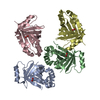
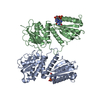
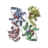
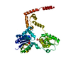
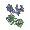
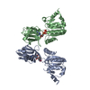
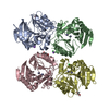
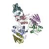
- PDB-4g0h: Crystal structure of the N-terminal domain of Helicobacter pylori... -
+ Open data

- Basic information
| Entry | Database: PDB / ID: 4g0h | ||||||
|---|---|---|---|---|---|---|---|
| Title | Crystal structure of the N-terminal domain of Helicobacter pylori CagA protein | ||||||
 Components Components | Cytotoxicity-associated immunodominant antigen | ||||||
 Keywords Keywords | TOXIN / cytotoxin / integrin beta 1 / PROTEIN BINDING | ||||||
| Function / homology |  Function and homology information Function and homology informationsymbiont-mediated perturbation of host cell cycle progression / toxin transmembrane transporter activity / molecular adaptor activity Similarity search - Function | ||||||
| Biological species |   Helicobacter pylori (bacteria) Helicobacter pylori (bacteria) | ||||||
| Method |  X-RAY DIFFRACTION / SYNCHROTRON / SAD / Resolution: 3.6 Å X-RAY DIFFRACTION / SYNCHROTRON / SAD / Resolution: 3.6 Å | ||||||
 Authors Authors | Kaplan-Turkoz, B. / Remaut, H. / Dian, C. / Louche, A. / Terradot, L. | ||||||
 Citation Citation | Journal: Proc.Natl.Acad.Sci.USA / Year: 2012 Title: Structural insights into Helicobacter pylori oncoprotein CagA interaction with beta 1 integrin. Authors: Kaplan-Turkoz, B. / Jimenez-Soto, L.F. / Dian, C. / Ertl, C. / Remaut, H. / Louche, A. / Tosi, T. / Haas, R. / Terradot, L. | ||||||
| History |
|
- Structure visualization
| Structure viewer | Molecule: MolmilJmol/JSmol |
|---|
- Downloads & links
-Download
| PDBx/mmCIF format | 4g0h.cif.gz | 104.9 KB | Display | PDBx/mmCIF format |
|---|---|---|---|---|
| PDB format | pdb4g0h.ent.gz | 74.3 KB | Display | PDB format |
| PDBx/mmJSON format | 4g0h.json.gz | Tree view | PDBx/mmJSON format | |
| Others |  Other downloads Other downloads |
-Validation report
| Arichive directory | https://data.pdbj.org/pub/pdb/validation_reports/g0/4g0hftp://data.pdbj.org/pub/pdb/validation_reports/g0/4g0h | HTTPS FTP |
|---|
-Related structure data
| Similar structure data |
|---|


-Links
 PDBj
PDBj- Assembly
| Deposited unit | 
| ||||||||
|---|---|---|---|---|---|---|---|---|---|
| 1 |
| ||||||||
| Unit cell |
|
-Components
| #1: Protein | Mass: 103005.117 Da / Num. of mol.: 1 / Fragment: UNP residues 1 to 884 Source method: isolated from a genetically manipulated source Source: (gene. exp.) Helicobacter pylori (bacteria) / Strain: 26695 / Gene: cag26, cagA, cai, HP0547, HP_0547 / Production host: |
|---|
-Experimental details
-Experiment
| Experiment | Method: X-RAY DIFFRACTION / Number of used crystals: 1 |
|---|
- Sample preparation
| Crystal | Density Matthews: 2.85 Å3/Da / Density % sol: 56.84 % |
|---|---|
| Crystal grow | Temperature: 293 K / Method: vapor diffusion, hanging drop / pH: 7 Details: 20% Ethanol, Na Cacodylate 100mM pH7, NaI 15mM, VAPOR DIFFUSION, HANGING DROP, temperature 293K |
-Data collection
| Diffraction | Mean temperature: 100 K | |||||||||||||||||||||||||||||||||||||||||||||||||||||||||||||||||||||||||||||
|---|---|---|---|---|---|---|---|---|---|---|---|---|---|---|---|---|---|---|---|---|---|---|---|---|---|---|---|---|---|---|---|---|---|---|---|---|---|---|---|---|---|---|---|---|---|---|---|---|---|---|---|---|---|---|---|---|---|---|---|---|---|---|---|---|---|---|---|---|---|---|---|---|---|---|---|---|---|---|
| Diffraction source | Source: SYNCHROTRON / Site: ESRF  / Beamline: ID29 / Wavelength: 0.97627 Å / Beamline: ID29 / Wavelength: 0.97627 Å | |||||||||||||||||||||||||||||||||||||||||||||||||||||||||||||||||||||||||||||
| Detector | Type: PSI PILATUS 6M / Detector: PIXEL / Date: Apr 13, 2011 | |||||||||||||||||||||||||||||||||||||||||||||||||||||||||||||||||||||||||||||
| Radiation | Protocol: SINGLE WAVELENGTH / Monochromatic (M) / Laue (L): M / Scattering type: x-ray | |||||||||||||||||||||||||||||||||||||||||||||||||||||||||||||||||||||||||||||
| Radiation wavelength | Wavelength: 0.97627 Å / Relative weight: 1 | |||||||||||||||||||||||||||||||||||||||||||||||||||||||||||||||||||||||||||||
| Reflection | Redundancy: 6.1 % / Av σ(I) over netI: 2.3 / Number: 76085 / Rsym value: 0.07 / D res high: 3.8 Å / D res low: 91.256 Å / Num. obs: 12510 / % possible obs: 99.9 | |||||||||||||||||||||||||||||||||||||||||||||||||||||||||||||||||||||||||||||
| Diffraction reflection shell |
| |||||||||||||||||||||||||||||||||||||||||||||||||||||||||||||||||||||||||||||
| Reflection | Resolution: 3.6→90.874 Å / Num. all: 13408 / Num. obs: 13408 / % possible obs: 92.8 % / Observed criterion σ(F): 1 / Observed criterion σ(I): 1 / Redundancy: 3.4 % / Rsym value: 0.076 / Net I/σ(I): 6.2 | |||||||||||||||||||||||||||||||||||||||||||||||||||||||||||||||||||||||||||||
| Reflection shell |
|
- Processing
| Software |
| ||||||||||||||||||||||||||||||||||||||||||
|---|---|---|---|---|---|---|---|---|---|---|---|---|---|---|---|---|---|---|---|---|---|---|---|---|---|---|---|---|---|---|---|---|---|---|---|---|---|---|---|---|---|---|---|
| Refinement | Method to determine structure: SAD / Resolution: 3.6→62.752 Å / Occupancy max: 1 / Occupancy min: 1 / SU ML: 0.62 / σ(F): 1.37 / Phase error: 39.45 / Stereochemistry target values: ML
| ||||||||||||||||||||||||||||||||||||||||||
| Solvent computation | Shrinkage radii: 1.1 Å / VDW probe radii: 1.3 Å / Solvent model: FLAT BULK SOLVENT MODEL | ||||||||||||||||||||||||||||||||||||||||||
| Displacement parameters | Biso mean: 166.8307 Å2
| ||||||||||||||||||||||||||||||||||||||||||
| Refine analyze | Luzzati coordinate error obs: 0.51 Å | ||||||||||||||||||||||||||||||||||||||||||
| Refinement step | Cycle: LAST / Resolution: 3.6→62.752 Å
| ||||||||||||||||||||||||||||||||||||||||||
| Refine LS restraints |
| ||||||||||||||||||||||||||||||||||||||||||
| LS refinement shell |
|
