 Movie
Movie Controller

[English] 日本語
 Yorodumi
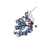
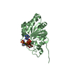
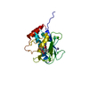
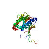
- PDB-1pt9: Crystal Structure Analysis of the DIII Component of Transhydrogen... -
+ Open data

- Basic information
| Entry | Database: PDB / ID: 1pt9 | ||||||
|---|---|---|---|---|---|---|---|
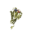
| Title | Crystal Structure Analysis of the DIII Component of Transhydrogenase with a Thio-Nicotinamide Nucleotide Analogue | ||||||
 Components Components | NAD(P) transhydrogenase, mitochondrial | ||||||
 Keywords Keywords | OXIDOREDUCTASE / Transhydrogenase / thio-nicotinamide / mitochondria / proton translocation | ||||||
| Function / homology |  Function and homology information Function and homology informationresponse to vitamin / NAD(P)+ transhydrogenase (Si-specific) activity / proton-translocating NAD(P)+ transhydrogenase activity / proton-translocating NAD(P)+ transhydrogenase / Citric acid cycle (TCA cycle) / NADPH regeneration / intracellular oxygen homeostasis / respiratory chain complex / positive regulation of hydrogen peroxide catabolic process / positive regulation of mitochondrial membrane potential ...response to vitamin / NAD(P)+ transhydrogenase (Si-specific) activity / proton-translocating NAD(P)+ transhydrogenase activity / proton-translocating NAD(P)+ transhydrogenase / Citric acid cycle (TCA cycle) / NADPH regeneration / intracellular oxygen homeostasis / respiratory chain complex / positive regulation of hydrogen peroxide catabolic process / positive regulation of mitochondrial membrane potential / cellular oxidant detoxification / tricarboxylic acid cycle / reactive oxygen species metabolic process / cell redox homeostasis / proton transmembrane transport / NAD binding / NADP binding / mitochondrial inner membrane / negative regulation of apoptotic process / mitochondrion / membrane Similarity search - Function | ||||||
| Biological species |  Homo sapiens (human) Homo sapiens (human) | ||||||
| Method |  X-RAY DIFFRACTION / SYNCHROTRON / MOLECULAR REPLACEMENT / Resolution: 2.42 Å X-RAY DIFFRACTION / SYNCHROTRON / MOLECULAR REPLACEMENT / Resolution: 2.42 Å | ||||||
 Authors Authors | Singh, A. / Venning, J.D. / Quirk, P.G. / van Boxel, G.I. / Rodrigues, D.J. / White, S.A. / Jackson, J.B. | ||||||
 Citation Citation | Journal: J.Biol.Chem. / Year: 2003 Title: Interactions between transhydrogenase and thio-nicotinamide analogues of NAD(H) and NADP(H) underline the importance of nucleotide conformational changes in coupling to proton translocation Authors: Singh, A. / Venning, J.D. / Quirk, P.G. / Van Boxel, G.I. / Rodrigues, D.J. / White, S.A. / Jackson, J.B. | ||||||
| History |
|
- Structure visualization
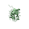
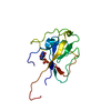
| Structure viewer | Molecule: MolmilJmol/JSmol |
|---|
- Downloads & links
-Download
| PDBx/mmCIF format | 1pt9.cif.gz | 88.4 KB | Display | PDBx/mmCIF format |
|---|---|---|---|---|
| PDB format | pdb1pt9.ent.gz | 66.8 KB | Display | PDB format |
| PDBx/mmJSON format | 1pt9.json.gz | Tree view | PDBx/mmJSON format | |
| Others |  Other downloads Other downloads |
-Validation report
| Arichive directory | https://data.pdbj.org/pub/pdb/validation_reports/pt/1pt9ftp://data.pdbj.org/pub/pdb/validation_reports/pt/1pt9 | HTTPS FTP |
|---|
-Related structure data
| Related structure data |  1ptjC  1djlS C: citing same article ( S: Starting model for refinement |
|---|---|
| Similar structure data |


-Links
 PDBj
PDBj

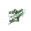
- Assembly
| Deposited unit | 
| ||||||||||||
|---|---|---|---|---|---|---|---|---|---|---|---|---|---|
| 1 | 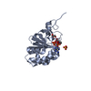
| ||||||||||||
| 2 | 
| ||||||||||||
| 3 | 
| ||||||||||||
| 4 | 
| ||||||||||||
| 5 | 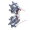
| ||||||||||||
| 6 | 
| ||||||||||||
| Unit cell |
| ||||||||||||
| Components on special symmetry positions |
|
-Components
| #1: Protein | Mass: 22281.652 Da / Num. of mol.: 2 / Fragment: residues 880-1086 / Source method: isolated from a natural source / Source: (natural) Homo sapiens (human) / Organ: Heart / References: UniProt: Q13423, EC: 1.6.1.2#2: Chemical |   Mass: 96.063 Da / Num. of mol.: 2 / Source method: obtained synthetically / Formula: SO4 Mass: 96.063 Da / Num. of mol.: 2 / Source method: obtained synthetically / Formula: SO4#3: Chemical | 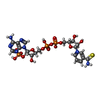  Mass: 759.471 Da / Num. of mol.: 2 / Source method: obtained synthetically / Formula: C21H28N7O16P3S Mass: 759.471 Da / Num. of mol.: 2 / Source method: obtained synthetically / Formula: C21H28N7O16P3S#4: Chemical |   Mass: 92.094 Da / Num. of mol.: 2 / Source method: obtained synthetically / Formula: C3H8O3 Mass: 92.094 Da / Num. of mol.: 2 / Source method: obtained synthetically / Formula: C3H8O3#5: Water | ChemComp-HOH / |  Mass: 18.015 Da / Num. of mol.: 171 / Source method: isolated from a natural source / Formula: H2O Mass: 18.015 Da / Num. of mol.: 171 / Source method: isolated from a natural source / Formula: H2O |
|---|
-Experimental details
-Experiment
| Experiment | Method: X-RAY DIFFRACTION / Number of used crystals: 1 |
|---|
- Sample preparation
| Crystal | Density Matthews: 2.32 Å3/Da / Density % sol: 46.98 % |
|---|---|
| Crystal grow | Method: vapor diffusion, sitting drop / pH: 7 Details: ammonium sulfate, peg 400, glycerol, pH 7.00, VAPOR DIFFUSION, SITTING DROP, temperature 100K |
| Crystal grow | *PLUS Method: vapor diffusion, hanging dropDetails: unpublished data, White, S.A., (2000) Structure Fold.Des., 8, 1. |
-Data collection
| Diffraction | Mean temperature: 100 K |
|---|---|
| Diffraction source | Source: SYNCHROTRON / Site: ESRF  / Beamline: ID14-1 / Beamline: ID14-1 |
| Detector | Detector: CCD |
| Radiation | Protocol: SINGLE WAVELENGTH / Monochromatic (M) / Laue (L): M / Scattering type: x-ray |
| Radiation wavelength | Relative weight: 1 |
| Reflection | Resolution: 2.42→36.82 Å / Num. all: 102352 / Num. obs: 100817 / % possible obs: 98.5 % / Biso Wilson estimate: 50.91 Å2 / Rsym value: 0.077 / Net I/σ(I): 6.8 |
| Reflection shell | Resolution: 2.42→2.55 Å / Mean I/σ(I) obs: 2.2 / Num. unique all: 2312 / Rsym value: 0.319 / % possible all: 96.2 |
| Reflection | *PLUS Num. obs: 16825 / Redundancy: 6 % / Num. measured all: 100817 / Rmerge(I) obs: 0.077 |
| Reflection shell | *PLUS % possible obs: 96.2 % / Redundancy: 4.9 % / Num. unique obs: 2312 / Num. measured obs: 11365 / Rmerge(I) obs: 0.319 |
- Processing
| Software |
| ||||||||||||||||||||||||||||
|---|---|---|---|---|---|---|---|---|---|---|---|---|---|---|---|---|---|---|---|---|---|---|---|---|---|---|---|---|---|
| Refinement | Method to determine structure: MOLECULAR REPLACEMENT Starting model: PDB ENTRY 1DJL Resolution: 2.42→36.8 Å / Isotropic thermal model: Isotropic / Cross valid method: THROUGHOUT / Stereochemistry target values: Engh & Huber
| ||||||||||||||||||||||||||||
| Displacement parameters | Biso mean: 38.71 Å2 | ||||||||||||||||||||||||||||
| Refinement step | Cycle: LAST / Resolution: 2.42→36.8 Å
| ||||||||||||||||||||||||||||
| Refine LS restraints |
| ||||||||||||||||||||||||||||
| LS refinement shell | Resolution: 2.42→2.45 Å
| ||||||||||||||||||||||||||||
| Xplor file |
| ||||||||||||||||||||||||||||
| Refinement | *PLUS Lowest resolution: 36.82 Å / % reflection Rfree: 5 % / Rfactor Rfree: 0.278 / Rfactor Rwork: 0.2187 | ||||||||||||||||||||||||||||
| Solvent computation | *PLUS | ||||||||||||||||||||||||||||
| Displacement parameters | *PLUS |
