 Movie
Movie Controller

[English] 日本語
 Yorodumi
- PDB-1djl: THE CRYSTAL STRUCTURE OF HUMAN TRANSHYDROGENASE DOMAIN III WITH B... -
+ Open data

- Basic information
| Entry | Database: PDB / ID: 1djl | ||||||
|---|---|---|---|---|---|---|---|
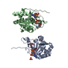
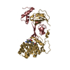
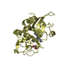
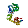
| Title | THE CRYSTAL STRUCTURE OF HUMAN TRANSHYDROGENASE DOMAIN III WITH BOUND NADP | ||||||
 Components Components | TRANSHYDROGENASE DIII | ||||||
 Keywords Keywords | OXIDOREDUCTASE / ROSSMANN FOLD DINUCLEOTIDE BINDING FOLD REVERSE BINDING OF NADP | ||||||
| Function / homology |  Function and homology information Function and homology informationresponse to vitamin / NAD(P)+ transhydrogenase (Si-specific) activity / proton-translocating NAD(P)+ transhydrogenase activity / proton-translocating NAD(P)+ transhydrogenase / Citric acid cycle (TCA cycle) / NADPH regeneration / intracellular oxygen homeostasis / respiratory chain complex / positive regulation of mitochondrial membrane potential / positive regulation of hydrogen peroxide catabolic process ...response to vitamin / NAD(P)+ transhydrogenase (Si-specific) activity / proton-translocating NAD(P)+ transhydrogenase activity / proton-translocating NAD(P)+ transhydrogenase / Citric acid cycle (TCA cycle) / NADPH regeneration / intracellular oxygen homeostasis / respiratory chain complex / positive regulation of mitochondrial membrane potential / positive regulation of hydrogen peroxide catabolic process / cellular oxidant detoxification / tricarboxylic acid cycle / reactive oxygen species metabolic process / proton transmembrane transport / cell redox homeostasis / NAD binding / NADP binding / mitochondrial inner membrane / negative regulation of apoptotic process / mitochondrion / membrane Similarity search - Function | ||||||
| Biological species |  Homo sapiens (human) Homo sapiens (human) | ||||||
| Method |  X-RAY DIFFRACTION / SYNCHROTRON / Resolution: 2 Å X-RAY DIFFRACTION / SYNCHROTRON / Resolution: 2 Å | ||||||
 Authors Authors | White, S.A. / Peak, S.J. / Cotton, N.P. / Jackson, J.B. | ||||||
 Citation Citation | Journal: Structure Fold.Des. / Year: 2000 Title: The high-resolution structure of the NADP(H)-binding component (dIII) of proton-translocating transhydrogenase from human heart mitochondria. Authors: White, S.A. / Peake, S.J. / McSweeney, S. / Leonard, G. / Cotton, N.P. / Jackson, J.B. #1: Journal: To be PublishedTitle: Structure and Mechanism of Proton-Translocating Transhydrogenase: A Mini-Review Authors: Jackson, J.B. / Peak, S.J. / White, S.A. #2: Journal: To be PublishedTitle: The NADPH-Binding Component (dIII) of Human-Heart Transhydrogenase: Crystallisation and Preliminary Crystallographic Analysis Authors: Peak, S.J. / Jackson, J.B. / White, S.A. | ||||||
| History |
|
- Structure visualization
| Structure viewer | Molecule: MolmilJmol/JSmol |
|---|
- Downloads & links
-Download
| PDBx/mmCIF format | 1djl.cif.gz | 85.3 KB | Display | PDBx/mmCIF format |
|---|---|---|---|---|
| PDB format | pdb1djl.ent.gz | 65.1 KB | Display | PDB format |
| PDBx/mmJSON format | 1djl.json.gz | Tree view | PDBx/mmJSON format | |
| Others |  Other downloads Other downloads |
-Validation report
| Arichive directory | https://data.pdbj.org/pub/pdb/validation_reports/dj/1djlftp://data.pdbj.org/pub/pdb/validation_reports/dj/1djl | HTTPS FTP |
|---|
-Related structure data
| Similar structure data |
|---|


-Links
 PDBj
PDBj

- Assembly
| Deposited unit | 
| ||||||||
|---|---|---|---|---|---|---|---|---|---|
| 1 |
| ||||||||
| Unit cell |
|
-Components
| #1: Protein | Mass: 22281.652 Da / Num. of mol.: 2 / Fragment: RESIDUES 837-1086 Source method: isolated from a genetically manipulated source Source: (gene. exp.) Homo sapiens (human) / Organ: HEART / Production host:  References: UniProt: Q13423, NAD(P)+ transhydrogenase (Si-specific) #2: Chemical |   Mass: 96.063 Da / Num. of mol.: 2 / Source method: obtained synthetically / Formula: SO4 Mass: 96.063 Da / Num. of mol.: 2 / Source method: obtained synthetically / Formula: SO4#3: Chemical |   Mass: 743.405 Da / Num. of mol.: 2 / Source method: obtained synthetically / Formula: C21H28N7O17P3 Mass: 743.405 Da / Num. of mol.: 2 / Source method: obtained synthetically / Formula: C21H28N7O17P3#4: Chemical |   Mass: 92.094 Da / Num. of mol.: 2 / Source method: obtained synthetically / Formula: C3H8O3 Mass: 92.094 Da / Num. of mol.: 2 / Source method: obtained synthetically / Formula: C3H8O3#5: Water | ChemComp-HOH / |  Mass: 18.015 Da / Num. of mol.: 80 / Source method: isolated from a natural source / Formula: H2O Mass: 18.015 Da / Num. of mol.: 80 / Source method: isolated from a natural source / Formula: H2O |
|---|
-Experimental details
-Experiment
| Experiment | Method: X-RAY DIFFRACTION / Number of used crystals: 2 |
|---|
- Sample preparation
| Crystal | Density Matthews: 2.37 Å3/Da / Density % sol: 48.18 % |
|---|---|
| Crystal grow | Temperature: 291 K / Method: vapor diffusion, hanging drop / pH: 7 Details: AMMONIUM SULPHATE, MOPS, DIOXANE, pH 7.0, VAPOR DIFFUSION, HANGING DROP, temperature 291K |
| Crystal grow | *PLUS Method: vapor diffusion, hanging dropDetails: SJ Peake, JB Jackeson and SA White, unpublished data |
-Data collection
| Diffraction | Mean temperature: 100 K |
|---|---|
| Diffraction source | Source: SYNCHROTRON / Site: ESRF  / Beamline: ID14-4 / Beamline: ID14-4 |
| Detector | Type: ADSC QUANTUM 4 / Detector: CCD |
| Radiation | Protocol: SINGLE WAVELENGTH / Monochromatic (M) / Laue (L): M / Scattering type: x-ray |
| Radiation wavelength | Relative weight: 1 |
- Processing
| Software |
| |||||||||||||||
|---|---|---|---|---|---|---|---|---|---|---|---|---|---|---|---|---|
| Refinement | Highest resolution: 2 Å /
| |||||||||||||||
| Refinement step | Cycle: LAST / Highest resolution: 2 Å
| |||||||||||||||
| Software | *PLUS Name: CNS / Classification: refinement | |||||||||||||||
| Refine LS restraints | *PLUS
|
