 Movie
Movie Controller


+ Open data

- Basic information
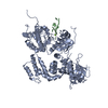
| Entry | Database: PDB / ID: 5w8s | ||||||||||||
|---|---|---|---|---|---|---|---|---|---|---|---|---|---|
| Title | Lipid A Disaccharide Synthase (LpxB)-7 solubilizing mutations | ||||||||||||
 Components Components | Lipid-A-disaccharide synthase | ||||||||||||
 Keywords Keywords | TRANSFERASE / glycosyltransferase B / Rossmann-like / C-terminal swap / dimer / lipid A disaccharide synthase / Raetz pathway / lipid A synthesis pathway / lipiopolysaccharide synthesis | ||||||||||||
| Function / homology |  Function and homology information Function and homology informationlipid-A-disaccharide synthase / lipid-A-disaccharide synthase activity / extrinsic component of plasma membrane / extrinsic component of cytoplasmic side of plasma membrane / lipid A biosynthetic process / phospholipid binding / membrane / identical protein binding / cytoplasm Similarity search - Function | ||||||||||||
| Biological species |  | ||||||||||||
| Method |  X-RAY DIFFRACTION / SYNCHROTRON / SAD / Resolution: 2.1 Å X-RAY DIFFRACTION / SYNCHROTRON / SAD / Resolution: 2.1 Å | ||||||||||||
 Authors Authors | Bohl, T.E. / Aihara, H. / Shi, K. / Lee, J.K. | ||||||||||||
| Funding support |  United States, 3items United States, 3items
| ||||||||||||
 Citation Citation | Journal: Nat Commun / Year: 2018 Title: Crystal structure of lipid A disaccharide synthase LpxB from Escherichia coli. Authors: Bohl, T.E. / Shi, K. / Lee, J.K. / Aihara, H. | ||||||||||||
| History |
|
- Structure visualization
| Structure viewer | Molecule: MolmilJmol/JSmol |
|---|
- Downloads & links
-Download
| PDBx/mmCIF format | 5w8s.cif.gz | 222.1 KB | Display | PDBx/mmCIF format |
|---|---|---|---|---|
| PDB format | pdb5w8s.ent.gz | 178.6 KB | Display | PDB format |
| PDBx/mmJSON format | 5w8s.json.gz | Tree view | PDBx/mmJSON format | |
| Others |  Other downloads Other downloads |
-Validation report
| Arichive directory | https://data.pdbj.org/pub/pdb/validation_reports/w8/5w8sftp://data.pdbj.org/pub/pdb/validation_reports/w8/5w8s | HTTPS FTP |
|---|
-Related structure data


-Links
 PDBj
PDBj
- Assembly
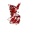
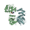
| Deposited unit | 
| ||||||||||||
|---|---|---|---|---|---|---|---|---|---|---|---|---|---|
| 1 | 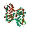
| ||||||||||||
| Unit cell |
| ||||||||||||
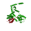
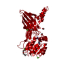
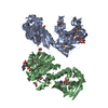
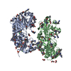
| Details | Enzymatic activity assay showed that LpxB with mutations in the swapped portion of the dimer can be complemented by LpxB with mutation in the unswapped portion by forming an LpxB mutant heterodimer with more activity than individual LpxB mutants. Thus, C-terminal swapped dimerization occurs and creates one intact active site in each LpxB mutant heterodimer. |
-Components
| #1: Protein | Mass: 42281.875 Da / Num. of mol.: 1 / Mutation: V66S, V68S, L69S, L72S, L75S, L76S, M207S Source method: isolated from a genetically manipulated source Source: (gene. exp.) Details (production host): cleavable His-tagged N-terminal SUMO Production host: References: UniProt: A0A140NAT1, UniProt: P10441*PLUS, lipid-A-disaccharide synthase | ||||
|---|---|---|---|---|---|
| #2: Chemical |   Mass: 22.990 Da / Num. of mol.: 2 / Source method: obtained synthetically / Formula: Na Mass: 22.990 Da / Num. of mol.: 2 / Source method: obtained synthetically / Formula: Na#3: Chemical | ChemComp-LI / |   Mass: 6.941 Da / Num. of mol.: 1 / Source method: obtained synthetically / Formula: Li Mass: 6.941 Da / Num. of mol.: 1 / Source method: obtained synthetically / Formula: Li#4: Water | ChemComp-HOH / |  Mass: 18.015 Da / Num. of mol.: 150 / Source method: isolated from a natural source / Formula: H2O Mass: 18.015 Da / Num. of mol.: 150 / Source method: isolated from a natural source / Formula: H2O |
-Experimental details
-Experiment
| Experiment | Method: X-RAY DIFFRACTION / Number of used crystals: 1 |
|---|
- Sample preparation
| Crystal | Density Matthews: 2.45 Å3/Da / Density % sol: 49.86 % / Description: bipyramidal |
|---|---|
| Crystal grow | Temperature: 292 K / Method: vapor diffusion, hanging drop / pH: 8.6 Details: 39% PEG 4000, 0.1 M Tris-HCl pH 8.6, 0.7 M LiCl mixed 1:1 with 8 g/L protein in 0.3 M NaCl, 5% glycerol, 20 mM Tris-HCl pH 7.4, 5 mM DTT with 100 mM trimethylammonium chloride additive used ...Details: 39% PEG 4000, 0.1 M Tris-HCl pH 8.6, 0.7 M LiCl mixed 1:1 with 8 g/L protein in 0.3 M NaCl, 5% glycerol, 20 mM Tris-HCl pH 7.4, 5 mM DTT with 100 mM trimethylammonium chloride additive used at 10 mM or 10% of drop volume |
-Data collection
| Diffraction | Mean temperature: 100 K |
|---|---|
| Diffraction source | Source: SYNCHROTRON / Site: APS / Beamline: 24-ID-E / Wavelength: 0.97918 Å |
| Detector | Type: ADSC QUANTUM 315 / Detector: CCD / Date: Jul 20, 2015 |
| Radiation | Protocol: SINGLE WAVELENGTH / Monochromatic (M) / Laue (L): M / Scattering type: x-ray |
| Radiation wavelength | Wavelength: 0.97918 Å / Relative weight: 1 |
| Reflection | Resolution: 2.1→155.14 Å / Num. obs: 24865 / % possible obs: 99.4 % / Redundancy: 3.5 % / Biso Wilson estimate: 40.03 Å2 / CC1/2: 0.998 / Rmerge(I) obs: 0.066 / Rpim(I) all: 0.041 / Net I/σ(I): 13.5 |
| Reflection shell | Resolution: 2.1→2.16 Å / Redundancy: 3.6 % / Rmerge(I) obs: 0.997 / Mean I/σ(I) obs: 1.6 / Num. unique obs: 2012 / CC1/2: 0.55 / Rpim(I) all: 0.615 / % possible all: 99.9 |
- Processing
| Software |
| ||||||||||||||||||
|---|---|---|---|---|---|---|---|---|---|---|---|---|---|---|---|---|---|---|---|
| Refinement | Method to determine structure: SAD / Resolution: 2.1→55.07 Å / SU ML: 0.3 / Cross valid method: FREE R-VALUE / Phase error: 23.86
| ||||||||||||||||||
| Solvent computation | Shrinkage radii: 0.9 Å / VDW probe radii: 1.11 Å | ||||||||||||||||||
| Displacement parameters | Biso mean: 45.6 Å2 | ||||||||||||||||||
| Refinement step | Cycle: LAST / Resolution: 2.1→55.07 Å
| ||||||||||||||||||
| Refine LS restraints | Type: GEOSTD + MON.LIB. + CDL v1.2 | ||||||||||||||||||
| LS refinement shell | Resolution: 2.1→2.15 Å
|
