 Movie
Movie Controller

[English] 日本語
 Yorodumi
- PDB-2etv: Crystal structure of a putative fe(iii) abc transporter (tm0189) ... -
+ Open data

- Basic information
| Entry | Database: PDB / ID: 2etv | ||||||
|---|---|---|---|---|---|---|---|
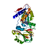
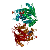
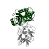
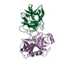
| Title | Crystal structure of a putative fe(iii) abc transporter (tm0189) from thermotoga maritima msb8 at 1.70 A resolution | ||||||
 Components Components | iron(III) ABC transporter, periplasmic iron-binding protein, putative | ||||||
 Keywords Keywords | METAL BINDING PROTEIN / Periplasmic iron-binding protein / structural genomics / Joint Center for Structural Genomics / JCSG / Protein Structure Initiative / PSI-2 | ||||||
| Function / homology |  Function and homology information Function and homology information: / ABC transporter periplasmic binding domain / Periplasmic binding protein / Iron siderophore/cobalamin periplasmic-binding domain profile. / Nitrogenase molybdenum iron protein domain / Rossmann fold / 3-Layer(aba) Sandwich / Alpha Beta Similarity search - Domain/homology | ||||||
| Biological species |   Thermotoga maritima (bacteria) Thermotoga maritima (bacteria) | ||||||
| Method |  X-RAY DIFFRACTION / SYNCHROTRON / MAD / Resolution: 1.7 Å X-RAY DIFFRACTION / SYNCHROTRON / MAD / Resolution: 1.7 Å | ||||||
 Authors Authors | Joint Center for Structural Genomics (JCSG) | ||||||
 Citation Citation | Journal: To be published Title: Crystal structure of IRON(III) ABC transporter, periplasmic iron-binding protein, putative (tm0189) from THERMOTOGA MARITIMA at 1.70 A resolution Authors: Joint Center for Structural Genomics (JCSG) | ||||||
| History |
| ||||||
| Remark 999 | SEQUENCE (1) THE CONSTRUCT EXPRESSED COMPRISED AN N-TERMINAL PURIFICATION TAG [MGSDKIHHHHHH] ...SEQUENCE (1) THE CONSTRUCT EXPRESSED COMPRISED AN N-TERMINAL PURIFICATION TAG [MGSDKIHHHHHH] FOLLOWED BY RESIDUES 25-358 OF THE PREDICTED TM0189 GENE PRODUCT. IN ORDER TO REMOVE A PREDICTED TRANSMEMBRANE HELIX, THE FIRST 24 RESIDUES WERE NOT INCLUDED IN THE CONSTRUCT. (2) THE PROTEIN WAS REDUCTIVELY METHYLATED PRIOR TO CRYSTALLIZATION. |
- Structure visualization
| Structure viewer | Molecule: MolmilJmol/JSmol |
|---|
- Downloads & links
-Download
| PDBx/mmCIF format | 2etv.cif.gz | 170 KB | Display | PDBx/mmCIF format |
|---|---|---|---|---|
| PDB format | pdb2etv.ent.gz | 138.7 KB | Display | PDB format |
| PDBx/mmJSON format | 2etv.json.gz | Tree view | PDBx/mmJSON format | |
| Others |  Other downloads Other downloads |
-Validation report
| Arichive directory | https://data.pdbj.org/pub/pdb/validation_reports/et/2etvftp://data.pdbj.org/pub/pdb/validation_reports/et/2etv | HTTPS FTP |
|---|
-Related structure data
| Similar structure data | |
|---|---|
| Other databases |

-Links
 PDBj
PDBj- Assembly
| Deposited unit | 
| ||||||||||||||||||
|---|---|---|---|---|---|---|---|---|---|---|---|---|---|---|---|---|---|---|---|
| 1 | 
| ||||||||||||||||||
| 2 | 
| ||||||||||||||||||
| 3 | 
| ||||||||||||||||||
| Unit cell |
| ||||||||||||||||||
| Components on special symmetry positions |
| ||||||||||||||||||
| Noncrystallographic symmetry (NCS) | NCS domain:
NCS domain segments: Component-ID: 1 / Ens-ID: 1 / Beg label comp-ID: HIS / End label comp-ID: TRP / Refine code: 2 / Auth seq-ID: -1 - 358 / Label seq-ID: 11 - 346
|
-Components
-Protein , 1 types, 2 molecules AB
| #1: Protein | Mass: 39628.863 Da / Num. of mol.: 2 Source method: isolated from a genetically manipulated source Source: (gene. exp.) Thermotoga maritima (bacteria) / Strain: MSB8 / Gene: tm0189 / Production host: |
|---|
-Non-polymers , 5 types, 801 molecules 


| #2: Chemical | ChemComp-NI /  Mass: 58.693 Da / Num. of mol.: 1 / Source method: obtained synthetically / Formula: Ni Mass: 58.693 Da / Num. of mol.: 1 / Source method: obtained synthetically / Formula: Ni | ||||||
|---|---|---|---|---|---|---|---|
| #3: Chemical |  Mass: 35.453 Da / Num. of mol.: 2 / Source method: obtained synthetically / Formula: Cl Mass: 35.453 Da / Num. of mol.: 2 / Source method: obtained synthetically / Formula: Cl#4: Chemical | ChemComp-PO4 / |  Mass: 94.971 Da / Num. of mol.: 1 / Source method: obtained synthetically / Formula: PO4 Mass: 94.971 Da / Num. of mol.: 1 / Source method: obtained synthetically / Formula: PO4#5: Chemical | ChemComp-EDO /  Mass: 62.068 Da / Num. of mol.: 19 / Source method: obtained synthetically / Formula: C2H6O2 Mass: 62.068 Da / Num. of mol.: 19 / Source method: obtained synthetically / Formula: C2H6O2#6: Water | ChemComp-HOH / | Mass: 18.015 Da / Num. of mol.: 778 / Source method: isolated from a natural source / Formula: H2O |
-Experimental details
-Experiment
| Experiment | Method: X-RAY DIFFRACTION / Number of used crystals: 1 |
|---|
- Sample preparation
| Crystal | Density Matthews: 2.6 Å3/Da / Density % sol: 51.8 % |
|---|---|
| Crystal grow | Temperature: 277 K / Method: vapor diffusion, sitting drop, nanodrop Details: 0.2M (NH4)2HPO4, 20.0% PEG-3350, No Buffer pH 7.9, VAPOR DIFFUSION,SITTING DROP,NANODROP, temperature 277K |
-Data collection
| Diffraction | Mean temperature: 100 K | |||||||||||||||||||||||||||||||||||||||||||||||||||||||||||||||||||||||||||||
|---|---|---|---|---|---|---|---|---|---|---|---|---|---|---|---|---|---|---|---|---|---|---|---|---|---|---|---|---|---|---|---|---|---|---|---|---|---|---|---|---|---|---|---|---|---|---|---|---|---|---|---|---|---|---|---|---|---|---|---|---|---|---|---|---|---|---|---|---|---|---|---|---|---|---|---|---|---|---|
| Diffraction source | Source: SYNCHROTRON / Site: APS  / Beamline: 23-ID-D / Wavelength: 0.97960, 0.95372 / Beamline: 23-ID-D / Wavelength: 0.97960, 0.95372 | |||||||||||||||||||||||||||||||||||||||||||||||||||||||||||||||||||||||||||||
| Detector | Type: MARMOSAIC 300 mm CCD / Detector: CCD / Date: Aug 4, 2005 / Details: Adjustable focusing mirrors in K-B geometry | |||||||||||||||||||||||||||||||||||||||||||||||||||||||||||||||||||||||||||||
| Radiation | Monochromator: DOUBLE CRYSTAL, SI(111) / Protocol: MAD / Monochromatic (M) / Laue (L): M / Scattering type: x-ray | |||||||||||||||||||||||||||||||||||||||||||||||||||||||||||||||||||||||||||||
| Radiation wavelength |
| |||||||||||||||||||||||||||||||||||||||||||||||||||||||||||||||||||||||||||||
| Reflection | Resolution: 1.7→30 Å / Num. obs: 92056 / % possible obs: 92.8 % / Redundancy: 5.65 % / Rmerge(I) obs: 0.103 / Net I/σ(I): 6.78 | |||||||||||||||||||||||||||||||||||||||||||||||||||||||||||||||||||||||||||||
| Reflection shell | Diffraction-ID: 1
|
-Phasing
| Phasing | Method: MAD |
|---|
- Processing
| Software |
| ||||||||||||||||||||||||||||||||||||||||||||||||||||||||||||||||||||||||||||||||||||||||||||||||||||||||||||||||||||||||||||||||||
|---|---|---|---|---|---|---|---|---|---|---|---|---|---|---|---|---|---|---|---|---|---|---|---|---|---|---|---|---|---|---|---|---|---|---|---|---|---|---|---|---|---|---|---|---|---|---|---|---|---|---|---|---|---|---|---|---|---|---|---|---|---|---|---|---|---|---|---|---|---|---|---|---|---|---|---|---|---|---|---|---|---|---|---|---|---|---|---|---|---|---|---|---|---|---|---|---|---|---|---|---|---|---|---|---|---|---|---|---|---|---|---|---|---|---|---|---|---|---|---|---|---|---|---|---|---|---|---|---|---|---|---|
| Refinement | Method to determine structure: MAD / Resolution: 1.7→30 Å / Cor.coef. Fo:Fc: 0.969 / Cor.coef. Fo:Fc free: 0.947 / SU B: 4.077 / SU ML: 0.071 / TLS residual ADP flag: LIKELY RESIDUAL / Cross valid method: THROUGHOUT / ESU R: 0.09 / ESU R Free: 0.097 Stereochemistry target values: MAXIMUM LIKELIHOOD WITH PHASES Details: 1) HYDROGENS HAVE BEEN ADDED IN THE RIDING POSITIONS. 2) A NICKEL ION IS COORDINATED IN AN OCTAHEDRAL COMPLEX TO THE SIDECHAINS OF HIS-1, HIS-3 ON THE SAME CHAIN; HIS-2, HIS-4 ON A SYMMETRY- ...Details: 1) HYDROGENS HAVE BEEN ADDED IN THE RIDING POSITIONS. 2) A NICKEL ION IS COORDINATED IN AN OCTAHEDRAL COMPLEX TO THE SIDECHAINS OF HIS-1, HIS-3 ON THE SAME CHAIN; HIS-2, HIS-4 ON A SYMMETRY-RELATED CHAIN, AND TWO WATER MOLECULES. X-RAY FLUORESCENCE SUPPORTS THE ASSIGNMENT OF THE NICKEL ION. 3) A MET-INHIBITION PROTOCOL WAS USED FOR SELENOMETHIONINE INCORPORATION DURING PROTEIN EXPRESSION. THE OCCUPANCY OF THE SE ATOMS IN THE MSE RESIDUES WAS REDUCED TO 0.75 TO ACCOUNT FOR THE REDUCED SCATTERING POWER DUE TO PARTIAL S-MET INCORPORATION. 4) LYSINES 304 APPEARS TO HAVE BEEN PROTECTED FROM REDUCTIVE METHYLATION AND WAS MODELED AS LYSINE IN BOTH CHAINS. ALL OTHER LYSINES HAVE BEEN MODELED AS MLY (N-DIMETHYL-LYSINE).
| ||||||||||||||||||||||||||||||||||||||||||||||||||||||||||||||||||||||||||||||||||||||||||||||||||||||||||||||||||||||||||||||||||
| Solvent computation | Ion probe radii: 0.8 Å / Shrinkage radii: 0.8 Å / VDW probe radii: 1.2 Å / Solvent model: MASK | ||||||||||||||||||||||||||||||||||||||||||||||||||||||||||||||||||||||||||||||||||||||||||||||||||||||||||||||||||||||||||||||||||
| Displacement parameters | Biso mean: 20.72 Å2
| ||||||||||||||||||||||||||||||||||||||||||||||||||||||||||||||||||||||||||||||||||||||||||||||||||||||||||||||||||||||||||||||||||
| Refinement step | Cycle: LAST / Resolution: 1.7→30 Å
| ||||||||||||||||||||||||||||||||||||||||||||||||||||||||||||||||||||||||||||||||||||||||||||||||||||||||||||||||||||||||||||||||||
| Refine LS restraints |
| ||||||||||||||||||||||||||||||||||||||||||||||||||||||||||||||||||||||||||||||||||||||||||||||||||||||||||||||||||||||||||||||||||
| Refine LS restraints NCS | Dom-ID: 1 / Auth asym-ID: A / Ens-ID: 1 / Refine-ID: X-RAY DIFFRACTION
| ||||||||||||||||||||||||||||||||||||||||||||||||||||||||||||||||||||||||||||||||||||||||||||||||||||||||||||||||||||||||||||||||||
| LS refinement shell | Resolution: 1.7→1.744 Å / Total num. of bins used: 20
| ||||||||||||||||||||||||||||||||||||||||||||||||||||||||||||||||||||||||||||||||||||||||||||||||||||||||||||||||||||||||||||||||||
| Refinement TLS params. | Method: refined / Refine-ID: X-RAY DIFFRACTION
| ||||||||||||||||||||||||||||||||||||||||||||||||||||||||||||||||||||||||||||||||||||||||||||||||||||||||||||||||||||||||||||||||||
| Refinement TLS group | Refine-ID: X-RAY DIFFRACTION / Selection: all
|
