 Movie
Movie Controller

[English] 日本語
 Yorodumi
- PDB-4fzn: Crystal structure of syringacin M mutant D232A from Pseudomonas s... -
+ Open data

- Basic information
| Entry | Database: PDB / ID: 4fzn | ||||||
|---|---|---|---|---|---|---|---|
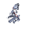
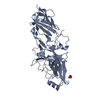
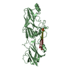
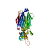
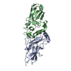
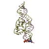
| Title | Crystal structure of syringacin M mutant D232A from Pseudomonas syringae pv. tomato DC3000 | ||||||
 Components Components | Bacteriocin | ||||||
 Keywords Keywords | ANTIMICROBIAL PROTEIN / Phosphatase | ||||||
| Function / homology |  Function and homology information Function and homology information | ||||||
| Biological species |  Pseudomonas syringae pv. tomato (bacteria) Pseudomonas syringae pv. tomato (bacteria) | ||||||
| Method |  X-RAY DIFFRACTION / SYNCHROTRON / MOLECULAR REPLACEMENT / Resolution: 3.12 Å X-RAY DIFFRACTION / SYNCHROTRON / MOLECULAR REPLACEMENT / Resolution: 3.12 Å | ||||||
 Authors Authors | Roszak, A.W. / Grinter, R. / Cogdell, J.R. / Walker, D. | ||||||
 Citation Citation | Journal: J.Biol.Chem. / Year: 2012 Title: The Crystal Structure of the Lipid II-degrading Bacteriocin Syringacin M Suggests Unexpected Evolutionary Relationships between Colicin M-like Bacteriocins. Authors: Grinter, R. / Roszak, A.W. / Cogdell, R.J. / Milner, J.J. / Walker, D. | ||||||
| History |
|
- Structure visualization
| Structure viewer | Molecule: MolmilJmol/JSmol |
|---|
- Downloads & links
-Download
| PDBx/mmCIF format | 4fzn.cif.gz | 122.6 KB | Display | PDBx/mmCIF format |
|---|---|---|---|---|
| PDB format | pdb4fzn.ent.gz | 97.1 KB | Display | PDB format |
| PDBx/mmJSON format | 4fzn.json.gz | Tree view | PDBx/mmJSON format | |
| Others |  Other downloads Other downloads |
-Validation report
| Arichive directory | https://data.pdbj.org/pub/pdb/validation_reports/fz/4fznftp://data.pdbj.org/pub/pdb/validation_reports/fz/4fzn | HTTPS FTP |
|---|
-Related structure data
| Related structure data | 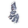 4fzlC 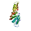 4fzmSC C: citing same article ( S: Starting model for refinement |
|---|---|
| Similar structure data |


-Links
 PDBj
PDBj- Assembly
| Deposited unit | 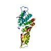
| ||||||||
|---|---|---|---|---|---|---|---|---|---|
| 1 |
| ||||||||
| 2 | 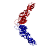
| ||||||||
| Unit cell |
|
-Components
| #1: Protein | Mass: 30539.068 Da / Num. of mol.: 1 / Mutation: D232A Source method: isolated from a genetically manipulated source Source: (gene. exp.) Pseudomonas syringae pv. tomato (bacteria)Strain: DC3000 / Gene: PSPTO_0572 / Plasmid: pETMDC / Production host: |
|---|---|
| #2: Water | ChemComp-HOH /  Mass: 18.015 Da / Num. of mol.: 27 / Source method: isolated from a natural source / Formula: H2O Mass: 18.015 Da / Num. of mol.: 27 / Source method: isolated from a natural source / Formula: H2O |
-Experimental details
-Experiment
| Experiment | Method: X-RAY DIFFRACTION / Number of used crystals: 1 |
|---|
- Sample preparation
| Crystal | Density % sol: 79.91 % |
|---|---|
| Crystal grow | Temperature: 289 K / Method: vapor diffusion, sitting drop / pH: 8.5 Details: 7% w/v PEG 8000, 30% v/v ethylene glycol 0.03 M CaCl2, 0.03 M MgCl2, 5% dimethyl sulfoxide, 0.1 M Bicine/Tris base, pH 8.5, VAPOR DIFFUSION, SITTING DROP, temperature 289K |
-Data collection
| Diffraction | Mean temperature: 100 K |
|---|---|
| Diffraction source | Source: SYNCHROTRON / Site: Diamond  / Beamline: I04 / Wavelength: 0.9795 Å / Beamline: I04 / Wavelength: 0.9795 Å |
| Detector | Type: ADSC QUANTUM 315r / Detector: CCD / Date: Nov 29, 2011 / Details: mirrors |
| Radiation | Monochromator: Si 111 channel / Protocol: SINGLE WAVELENGTH / Monochromatic (M) / Laue (L): M / Scattering type: x-ray |
| Radiation wavelength | Wavelength: 0.9795 Å / Relative weight: 1 |
| Reflection | Resolution: 3.12→40.09 Å / Num. all: 13347 / Num. obs: 13347 / % possible obs: 99.53 % / Observed criterion σ(F): 0 / Observed criterion σ(I): 0 / Redundancy: 15.5 % / Biso Wilson estimate: 107.9 Å2 / Rmerge(I) obs: 0.048 / Net I/σ(I): 26.9 |
| Reflection shell | Resolution: 3.12→3.2 Å / Redundancy: 16.2 % / Rmerge(I) obs: 0.787 / Mean I/σ(I) obs: 4.3 / Num. unique all: 1018 / % possible all: 100 |
- Processing
| Software |
| ||||||||||||||||||||||||||||||||||||||||||||||||||||||||||||
|---|---|---|---|---|---|---|---|---|---|---|---|---|---|---|---|---|---|---|---|---|---|---|---|---|---|---|---|---|---|---|---|---|---|---|---|---|---|---|---|---|---|---|---|---|---|---|---|---|---|---|---|---|---|---|---|---|---|---|---|---|---|
| Refinement | Method to determine structure: MOLECULAR REPLACEMENT Starting model: PDB ENTRY 4FZM Resolution: 3.12→40.09 Å / Cor.coef. Fo:Fc: 0.958 / Cor.coef. Fo:Fc free: 0.931 / SU B: 26.446 / SU ML: 0.209 / Isotropic thermal model: isotropic / Cross valid method: THROUGHOUT / ESU R: 0.383 / ESU R Free: 0.283 / Stereochemistry target values: MAXIMUM LIKELIHOOD / Details: HYDROGENS HAVE BEEN ADDED IN THE RIDING POSITIONS
| ||||||||||||||||||||||||||||||||||||||||||||||||||||||||||||
| Solvent computation | Ion probe radii: 0.8 Å / Shrinkage radii: 0.8 Å / VDW probe radii: 1.2 Å / Solvent model: MASK | ||||||||||||||||||||||||||||||||||||||||||||||||||||||||||||
| Displacement parameters | Biso mean: 98.702 Å2
| ||||||||||||||||||||||||||||||||||||||||||||||||||||||||||||
| Refinement step | Cycle: LAST / Resolution: 3.12→40.09 Å
| ||||||||||||||||||||||||||||||||||||||||||||||||||||||||||||
| Refine LS restraints |
| ||||||||||||||||||||||||||||||||||||||||||||||||||||||||||||
| LS refinement shell | Resolution: 3.121→3.201 Å / Total num. of bins used: 20
| ||||||||||||||||||||||||||||||||||||||||||||||||||||||||||||
| Refinement TLS params. | Method: refined / Origin x: -70.2903 Å / Origin y: 16.4942 Å / Origin z: -10.244 Å
|
