 Movie
Movie Controller


+ Open data

- Basic information
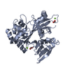
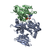
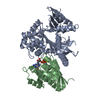
| Entry | Database: PDB / ID: 4fmb | ||||||
|---|---|---|---|---|---|---|---|
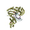
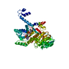
| Title | VirA-Rab1 complex structure | ||||||
 Components Components |
| ||||||
 Keywords Keywords | PROTEIN BINDING / alpha-beta fold / Rab1-GAP complex / Rab1 | ||||||
| Function / homology |  Function and homology information Function and homology informationpositive regulation of glycoprotein metabolic process / growth hormone secretion / COPII-coated vesicle cargo loading / RAB geranylgeranylation / melanosome transport / RAB GEFs exchange GTP for GDP on RABs / Golgi Cisternae Pericentriolar Stack Reorganization / vesicle transport along microtubule / COPII-mediated vesicle transport / COPI-dependent Golgi-to-ER retrograde traffic ...positive regulation of glycoprotein metabolic process / growth hormone secretion / COPII-coated vesicle cargo loading / RAB geranylgeranylation / melanosome transport / RAB GEFs exchange GTP for GDP on RABs / Golgi Cisternae Pericentriolar Stack Reorganization / vesicle transport along microtubule / COPII-mediated vesicle transport / COPI-dependent Golgi-to-ER retrograde traffic / virion assembly / transport vesicle membrane / Golgi organization / endoplasmic reticulum to Golgi vesicle-mediated transport / autophagosome assembly / COPI-mediated anterograde transport / vesicle-mediated transport / endomembrane system / substrate adhesion-dependent cell spreading / positive regulation of interleukin-8 production / small monomeric GTPase / intracellular protein transport / autophagy / endocytosis / melanosome / cell migration / G protein activity / early endosome / Hydrolases; Acting on peptide bonds (peptidases); Cysteine endopeptidases / defense response to bacterium / cadherin binding / Golgi membrane / cysteine-type endopeptidase activity / GTPase activity / GTP binding / Golgi apparatus / endoplasmic reticulum / proteolysis / extracellular exosome / extracellular region / metal ion binding / cytosol Similarity search - Function | ||||||
| Biological species |  Shigella flexneri (bacteria) Shigella flexneri (bacteria) Homo sapiens (human) Homo sapiens (human) | ||||||
| Method |  X-RAY DIFFRACTION / SYNCHROTRON / MOLECULAR REPLACEMENT / Resolution: 3.2 Å X-RAY DIFFRACTION / SYNCHROTRON / MOLECULAR REPLACEMENT / Resolution: 3.2 Å | ||||||
 Authors Authors | Shao, F. / Zhu, Y. | ||||||
 Citation Citation | Journal: Cell(Cambridge,Mass.) / Year: 2012 Title: Structurally Distinct Bacterial TBC-like GAPs Link Arf GTPase to Rab1 Inactivation to Counteract Host Defenses. Authors: Dong, N. / Zhu, Y. / Lu, Q. / Hu, L. / Zheng, Y. / Shao, F. | ||||||
| History |
|
- Structure visualization
| Structure viewer | Molecule: MolmilJmol/JSmol |
|---|
- Downloads & links
-Download
| PDBx/mmCIF format | 4fmb.cif.gz | 314.5 KB | Display | PDBx/mmCIF format |
|---|---|---|---|---|
| PDB format | pdb4fmb.ent.gz | 252.1 KB | Display | PDB format |
| PDBx/mmJSON format | 4fmb.json.gz | Tree view | PDBx/mmJSON format | |
| Others |  Other downloads Other downloads |
-Validation report
| Arichive directory | https://data.pdbj.org/pub/pdb/validation_reports/fm/4fmbftp://data.pdbj.org/pub/pdb/validation_reports/fm/4fmb | HTTPS FTP |
|---|
-Related structure data


-Links
 PDBj
PDBj


- Assembly
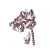
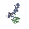
| Deposited unit | 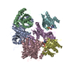
| ||||||||
|---|---|---|---|---|---|---|---|---|---|
| 1 | 
| ||||||||
| 2 | 
| ||||||||
| 3 | 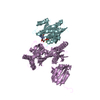
| ||||||||
| 4 | 
| ||||||||
| Unit cell |
|
-Components
-Protein , 2 types, 6 molecules ACEBDF
| #1: Protein | Mass: 40140.234 Da / Num. of mol.: 3 Source method: isolated from a genetically manipulated source Source: (gene. exp.) Shigella flexneri (bacteria) / Strain: 301 / Gene: CP0181, pWR501_0191, virA / Plasmid: pGEX-6p-2 / Production host: References: UniProt: Q7BU69, Hydrolases; Acting on peptide bonds (peptidases); Cysteine endopeptidases #2: Protein | Mass: 19384.988 Da / Num. of mol.: 3 Source method: isolated from a genetically manipulated source Source: (gene. exp.) Homo sapiens (human) / Gene: RAB1, RAB1A / Plasmid: PET28a / Production host: |
|---|
-Non-polymers , 4 types, 18 molecules 


| #3: Chemical |  Mass: 83.977 Da / Num. of mol.: 3 / Source method: obtained synthetically / Formula: AlF3 Mass: 83.977 Da / Num. of mol.: 3 / Source method: obtained synthetically / Formula: AlF3#4: Chemical |  Type: RNA linking / Mass: 443.201 Da / Num. of mol.: 3 / Source method: obtained synthetically / Formula: C10H15N5O11P2 / Comment: GDP, energy-carrying molecule*YM Type: RNA linking / Mass: 443.201 Da / Num. of mol.: 3 / Source method: obtained synthetically / Formula: C10H15N5O11P2 / Comment: GDP, energy-carrying molecule*YM#5: Chemical |  Mass: 24.305 Da / Num. of mol.: 3 / Source method: obtained synthetically / Formula: Mg Mass: 24.305 Da / Num. of mol.: 3 / Source method: obtained synthetically / Formula: Mg#6: Water | ChemComp-HOH / | Mass: 18.015 Da / Num. of mol.: 9 / Source method: isolated from a natural source / Formula: H2O |
|---|
-Details
| Has protein modification | Y |
|---|---|
| Sequence details | RESIDUES WERE CONFIRMED BY GENE SEQUENCING |
-Experimental details
-Experiment
| Experiment | Method: X-RAY DIFFRACTION / Number of used crystals: 1 |
|---|
- Sample preparation
| Crystal | Density Matthews: 3.55 Å3/Da / Density % sol: 65.35 % |
|---|---|
| Crystal grow | Temperature: 298 K / pH: 7.6 Details: 0.8 M ammonium sulfate and 0.1 M Tris-HCl (pH 7.6), VAPOR DIFFUSION, HANGING DROP, temperature 298K |
-Data collection
| Diffraction | Mean temperature: 100 K |
|---|---|
| Diffraction source | Source: SYNCHROTRON / Site: APS  / Beamline: 24-ID-C / Wavelength: 0.97918 / Beamline: 24-ID-C / Wavelength: 0.97918 |
| Detector | Type: ADSC QUANTUM 315 / Detector: CCD / Date: Mar 12, 2011 |
| Radiation | Monochromator: SI 111 CHANNEL / Protocol: SINGLE WAVELENGTH / Monochromatic (M) / Laue (L): M / Scattering type: x-ray |
| Radiation wavelength | Wavelength: 0.97918 Å / Relative weight: 1 |
| Reflection | Resolution: 3.2→50 Å / Num. obs: 40511 / % possible obs: 99.9 % / Observed criterion σ(I): 1 / Redundancy: 5.6 % / Rmerge(I) obs: 0.091 / Net I/σ(I): 4 |
- Processing
| Software |
| ||||||||||||||||||||||||||||||||||||||||||||||||||||||||||||||||||||||||||||||||
|---|---|---|---|---|---|---|---|---|---|---|---|---|---|---|---|---|---|---|---|---|---|---|---|---|---|---|---|---|---|---|---|---|---|---|---|---|---|---|---|---|---|---|---|---|---|---|---|---|---|---|---|---|---|---|---|---|---|---|---|---|---|---|---|---|---|---|---|---|---|---|---|---|---|---|---|---|---|---|---|---|---|
| Refinement | Method to determine structure: MOLECULAR REPLACEMENT / Resolution: 3.2→50 Å / Occupancy max: 1 / Occupancy min: 1 / σ(F): 0
| ||||||||||||||||||||||||||||||||||||||||||||||||||||||||||||||||||||||||||||||||
| Solvent computation | Bsol: 55.06 Å2 | ||||||||||||||||||||||||||||||||||||||||||||||||||||||||||||||||||||||||||||||||
| Displacement parameters | Biso mean: 105.24 Å2
| ||||||||||||||||||||||||||||||||||||||||||||||||||||||||||||||||||||||||||||||||
| Refinement step | Cycle: LAST / Resolution: 3.2→50 Å
| ||||||||||||||||||||||||||||||||||||||||||||||||||||||||||||||||||||||||||||||||
| Refine LS restraints |
| ||||||||||||||||||||||||||||||||||||||||||||||||||||||||||||||||||||||||||||||||
| LS refinement shell | Resolution: 3.2→3.31 Å / Total num. of bins used: 10
| ||||||||||||||||||||||||||||||||||||||||||||||||||||||||||||||||||||||||||||||||
| Xplor file |
|
