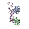
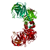
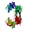
- PDB-4fgn: Crystal structure of the SV40 large T-antigen origin bining domai... -
+
Open data
ID or keywords:
Loading...
-
Basic information
Entry
Database: PDB / ID: 4fgn
Title
Crystal structure of the SV40 large T-antigen origin bining domain bound to Site I DNA
Components
(Site I DNA) x 2
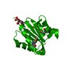
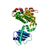
Large T antigen
Keywords
DNA BINDING PROTEIN/DNA / origin binding domain / DNA BINDING PROTEIN-DNA complex
Function / homology
Function and homology information
symbiont-mediated suppression of host JAK-STAT cascade via inhibition of JAK1 activity / bidirectional double-stranded viral DNA replication / viral DNA genome replication / DNA 3'-5' helicase / symbiont-mediated perturbation of host cell cycle G1/S transition checkpoint / DNA replication origin binding / helicase activity / single-stranded DNA binding / double-stranded DNA binding / symbiont-mediated perturbation of host ubiquitin-like protein modification ...symbiont-mediated suppression of host JAK-STAT cascade via inhibition of JAK1 activity / bidirectional double-stranded viral DNA replication / viral DNA genome replication / DNA 3'-5' helicase / symbiont-mediated perturbation of host cell cycle G1/S transition checkpoint / DNA replication origin binding / helicase activity / single-stranded DNA binding / double-stranded DNA binding / symbiont-mediated perturbation of host ubiquitin-like protein modification / DNA replication / symbiont-mediated suppression of host innate immune response / symbiont-mediated suppression of host type I interferon-mediated signaling pathway / host cell nucleus / ATP hydrolysis activity / zinc ion binding / ATP binding / identical protein binding Similarity search - Function
Replication Protein E1; Chain: A, - #20 / Replication Protein E1; Chain: A, / Large T antigen, polyomaviridae / Large T antigen, polyomavirus, C-terminal / Zinc finger, large T-antigen D1-type / Origin of replication binding protein / Polyomavirus large T antigen C-terminus / Large T-antigen (T-ag) origin-binding domain (OBD) profile. / Zinc finger large T-antigen (T-ag) D1-type profile. / T antigen, Ori-binding ...Replication Protein E1; Chain: A, - #20 / Replication Protein E1; Chain: A, / Large T antigen, polyomaviridae / Large T antigen, polyomavirus, C-terminal / Zinc finger, large T-antigen D1-type / Origin of replication binding protein / Polyomavirus large T antigen C-terminus / Large T-antigen (T-ag) origin-binding domain (OBD) profile. / Zinc finger large T-antigen (T-ag) D1-type profile. / T antigen, Ori-binding / Zinc finger, large T-antigen D1 domain superfamily / Helicase, superfamily 3, DNA virus / Superfamily 3 helicase of DNA viruses domain profile. / DnaJ molecular chaperone homology domain / dnaJ domain profile. / Chaperone J-domain superfamily / DnaJ domain / P-loop containing nucleoside triphosphate hydrolase / 3-Layer(aba) Sandwich / Alpha Beta Similarity search - Domain/homology
Mass: 15240.476 Da / Num. of mol.: 2 / Fragment: origin binding domain, UNP residues 131-260 Source method: isolated from a genetically manipulated source Source: (gene. exp.) Simian virus 40 / Production host: Escherichia coli (E. coli) / Strain (production host): Bl21 (DE3) References: UniProt: P03070, Hydrolases; Acting on acid anhydrides; Acting on acid anhydrides to facilitate cellular and subcellular movement
#2: DNA chain
SiteIDNA
Mass: 6963.528 Da / Num. of mol.: 1 / Source method: obtained synthetically / Details: This DNA sequence is termed Site 1 in SV40 / Source: (synth.) Simian virus 40
#3: DNA chain
SiteIDNA
Mass: 7158.603 Da / Num. of mol.: 1 / Source method: obtained synthetically / Details: This DNA sequence is from sv40 site I / Source: (synth.) Simian virus 40
Resolution: 3.2→47.99 Å / Cor.coef. Fo:Fc: 0.971 / Cor.coef. Fo:Fc free: 0.952 / SU B: 73.091 / SU ML: 0.517 / Cross valid method: THROUGHOUT / ESU R Free: 0.504 / Stereochemistry target values: MAXIMUM LIKELIHOOD / Details: HYDROGENS HAVE BEEN USED IF PRESENT IN THE INPUT
Rfactor
Num. reflection
% reflection
Selection details
Rfree
0.267
444
4.8 %
RANDOM
Rwork
0.188
-
-
-
obs
0.191
8841
99.8 %
-
all
-
9305
-
-
Solvent computation
Ion probe radii: 0.8 Å / Shrinkage radii: 0.8 Å / VDW probe radii: 1.2 Å / Solvent model: MASK
Displacement parameters
Biso mean: 137.11 Å2
Baniso -1
Baniso -2
Baniso -3
1-
-0.22 Å2
0 Å2
0 Å2
2-
-
-0.13 Å2
0 Å2
3-
-
-
0.35 Å2
Refinement step
Cycle: LAST / Resolution: 3.2→47.99 Å
Protein
Nucleic acid
Ligand
Solvent
Total
Num. atoms
2027
937
0
3
2967
Refine LS restraints
Refine-ID
Type
Dev ideal
Dev ideal target
Number
X-RAY DIFFRACTION
r_bond_refined_d
0.011
0.017
3132
X-RAY DIFFRACTION
r_bond_other_d
X-RAY DIFFRACTION
r_angle_refined_deg
1.767
1.737
4434
X-RAY DIFFRACTION
r_angle_other_deg
X-RAY DIFFRACTION
r_dihedral_angle_1_deg
8.224
5
248
X-RAY DIFFRACTION
r_dihedral_angle_2_deg
35.384
23.061
98
X-RAY DIFFRACTION
r_dihedral_angle_3_deg
20.75
15
345
X-RAY DIFFRACTION
r_dihedral_angle_4_deg
26.134
15
10
X-RAY DIFFRACTION
r_chiral_restr
0.102
0.2
448
X-RAY DIFFRACTION
r_gen_planes_refined
0.008
0.021
2062
X-RAY DIFFRACTION
r_gen_planes_other
X-RAY DIFFRACTION
r_nbd_refined
X-RAY DIFFRACTION
r_nbd_other
X-RAY DIFFRACTION
r_nbtor_refined
X-RAY DIFFRACTION
r_nbtor_other
X-RAY DIFFRACTION
r_xyhbond_nbd_refined
X-RAY DIFFRACTION
r_xyhbond_nbd_other
X-RAY DIFFRACTION
r_metal_ion_refined
X-RAY DIFFRACTION
r_metal_ion_other
X-RAY DIFFRACTION
r_symmetry_vdw_refined
X-RAY DIFFRACTION
r_symmetry_vdw_other
X-RAY DIFFRACTION
r_symmetry_hbond_refined
X-RAY DIFFRACTION
r_symmetry_hbond_other
X-RAY DIFFRACTION
r_symmetry_metal_ion_refined
X-RAY DIFFRACTION
r_symmetry_metal_ion_other
X-RAY DIFFRACTION
r_mcbond_it
X-RAY DIFFRACTION
r_mcbond_other
X-RAY DIFFRACTION
r_mcangle_it
X-RAY DIFFRACTION
r_scbond_it
X-RAY DIFFRACTION
r_scangle_it
X-RAY DIFFRACTION
r_rigid_bond_restr
X-RAY DIFFRACTION
r_sphericity_free
X-RAY DIFFRACTION
r_sphericity_bonded
LS refinement shell
Resolution: 3.2→3.28 Å / Total num. of bins used: 20
Rfactor
Num. reflection
% reflection
Rfree
0.266
29
-
Rwork
0.302
563
-
obs
-
-
97.69 %
Refinement TLS params.
Method: refined / Refine-ID: X-RAY DIFFRACTION
ID
L11 (°2)
L12 (°2)
L13 (°2)
L22 (°2)
L23 (°2)
L33 (°2)
S11 (Å °)
S12 (Å °)
S13 (Å °)
S21 (Å °)
S22 (Å °)
S23 (Å °)
S31 (Å °)
S32 (Å °)
S33 (Å °)
T11 (Å2)
T12 (Å2)
T13 (Å2)
T22 (Å2)
T23 (Å2)
T33 (Å2)
Origin x (Å)
Origin y (Å)
Origin z (Å)
1
4.3163
-2.8324
-1.0743
14.3332
2.3666
11.8625
0.0745
-0.2827
-0.9794
0.8123
-0.7172
0.6496
0.5555
-0.3503
0.6427
0.3479
-0.0302
0.2384
0.603
-0.0679
0.8232
1.5594
42.2545
-0.2878
2
10.8722
2.9635
1.3241
11.1175
0.2369
3.7342
0.9076
-0.6183
-0.5681
-0.2245
-0.7337
1.4421
1.6383
-1.9481
-0.1738
1.2112
-0.4074
-0.4374
1.5418
-0.1961
0.7115
3.1383
5.6793
-17.5268
3
4.5856
-5.0544
-0.7161
8.903
3.2412
3.2417
0.6374
0.3198
0.9739
-1.2909
-0.4605
-1.7934
-0.3216
0.3427
-0.1769
1.0534
0.2711
0.1618
0.6879
-0.0419
0.7649
17.6876
27.3756
-20.0936
4
2.5926
-2.6339
1.4469
4.0713
0.4706
4.1954
0.9031
0.5819
0.0307
-1.4441
-0.7736
0.2976
-0.2765
0.1121
-0.1295
0.9832
0.2006
0.2339
0.6251
-0.0533
0.7814
16.4447
29.9636
-20.6853
Refinement TLS group
ID
Refine-ID
Refine TLS-ID
Auth asym-ID
Auth seq-ID
1
X-RAY DIFFRACTION
1
A
133 - 258
2
X-RAY DIFFRACTION
2
B
134 - 257
3
X-RAY DIFFRACTION
3
Z
1 - 23
4
X-RAY DIFFRACTION
4
Y
1 - 23
+
About Yorodumi
-
News
-
Feb 9, 2022. New format data for meta-information of EMDB entries
New format data for meta-information of EMDB entries
Version 3 of the EMDB header file is now the official format.
The previous official version 1.9 will be removed from the archive.
In the structure databanks used in Yorodumi, some data are registered as the other names, "COVID-19 virus" and "2019-nCoV". Here are the details of the virus and the list of structure data.
Jan 31, 2019. EMDB accession codes are about to change! (news from PDBe EMDB page)
EMDB accession codes are about to change! (news from PDBe EMDB page)
The allocation of 4 digits for EMDB accession codes will soon come to an end. Whilst these codes will remain in use, new EMDB accession codes will include an additional digit and will expand incrementally as the available range of codes is exhausted. The current 4-digit format prefixed with “EMD-” (i.e. EMD-XXXX) will advance to a 5-digit format (i.e. EMD-XXXXX), and so on. It is currently estimated that the 4-digit codes will be depleted around Spring 2019, at which point the 5-digit format will come into force.
The EM Navigator/Yorodumi systems omit the EMD- prefix.
Related info.:Q: What is EMD? / ID/Accession-code notation in Yorodumi/EM Navigator
Yorodumi is a browser for structure data from EMDB, PDB, SASBDB, etc.
This page is also the successor to EM Navigator detail page, and also detail information page/front-end page for Omokage search.
The word "yorodu" (or yorozu) is an old Japanese word meaning "ten thousand". "mi" (miru) is to see.
Related info.:EMDB / PDB / SASBDB / Comparison of 3 databanks / Yorodumi Search / Aug 31, 2016. New EM Navigator & Yorodumi / Yorodumi Papers / Jmol/JSmol / Function and homology information / Changes in new EM Navigator and Yorodumi
 Movie
Movie Controller

 Yorodumi
 Open data
 Basic information
 Components
 Keywords
 Function and homology information
 Simian virus 40
 X-RAY DIFFRACTION /
  Authors
 Citation
 Structure visualization
 Downloads & links
 Other downloads
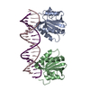


 PDBj
PDBj


 Assembly

 Mass: 18.015 Da / Num. of mol.: 3 / Source method: isolated from a natural source / Formula: H2O
 Sample preparation
 / Beamline: X29A / Wavelength: 0.9792
  Processing