 Movie
Movie Controller

[English] 日本語
 Yorodumi
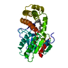
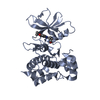
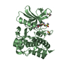
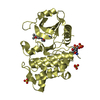
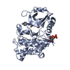
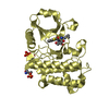
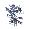
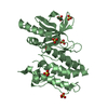
- PDB-3fve: Crystal structure of diaminopimelate epimerase Mycobacterium tube... -
+ Open data

- Basic information
| Entry | Database: PDB / ID: 3fve | ||||||
|---|---|---|---|---|---|---|---|
| Title | Crystal structure of diaminopimelate epimerase Mycobacterium tuberculosis DapF | ||||||
 Components Components | Diaminopimelate epimerase | ||||||
 Keywords Keywords | ISOMERASE / alpha/beta / Amino-acid biosynthesis / Lysine biosynthesis | ||||||
| Function / homology |  Function and homology information Function and homology informationdiaminopimelate epimerase / diaminopimelate epimerase activity / : / plasma membrane / cytosol Similarity search - Function | ||||||
| Biological species |   Mycobacterium tuberculosis (bacteria) Mycobacterium tuberculosis (bacteria) | ||||||
| Method |  X-RAY DIFFRACTION / SYNCHROTRON / MOLECULAR REPLACEMENT / molecular replacement / Resolution: 2.6 Å X-RAY DIFFRACTION / SYNCHROTRON / MOLECULAR REPLACEMENT / molecular replacement / Resolution: 2.6 Å | ||||||
 Authors Authors | Usha, V. / Dover, L.G. / Roper, D.I. / Futterer, K. / Besra, G.S. | ||||||
 Citation Citation | Journal: Acta Crystallogr.,Sect.D / Year: 2009 Title: Structure of the diaminopimelate epimerase DapF from Mycobacterium tuberculosis Authors: Usha, V. / Dover, L.G. / Roper, D.I. / Futterer, K. / Besra, G.S. | ||||||
| History |
|
- Structure visualization
| Structure viewer | Molecule: MolmilJmol/JSmol |
|---|
- Downloads & links
-Download
| PDBx/mmCIF format | 3fve.cif.gz | 65.5 KB | Display | PDBx/mmCIF format |
|---|---|---|---|---|
| PDB format | pdb3fve.ent.gz | 47.3 KB | Display | PDB format |
| PDBx/mmJSON format | 3fve.json.gz | Tree view | PDBx/mmJSON format | |
| Others |  Other downloads Other downloads |
-Validation report
| Arichive directory | https://data.pdbj.org/pub/pdb/validation_reports/fv/3fveftp://data.pdbj.org/pub/pdb/validation_reports/fv/3fve | HTTPS FTP |
|---|
-Related structure data
| Related structure data | 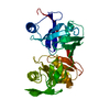 1bwzS S: Starting model for refinement |
|---|---|
| Similar structure data |


-Links
 PDBj
PDBj


- Assembly
| Deposited unit | 
| ||||||||
|---|---|---|---|---|---|---|---|---|---|
| 1 |
| ||||||||
| 2 | 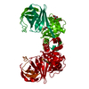
| ||||||||
| Unit cell |
| ||||||||
| Details | BIOLOGICAL UNIT IS THE SAME AS ASYM. |
-Components
| #1: Protein | Mass: 29864.635 Da / Num. of mol.: 1 Source method: isolated from a genetically manipulated source Source: (gene. exp.) Mycobacterium tuberculosis (bacteria) / Strain: H37RV / Gene: dapF, MT2798, MTCY154.06c, Rv2726c / Plasmid: pET28a / Production host: References: UniProt: P63897, UniProt: P9WP19*PLUS, diaminopimelate epimerase | ||
|---|---|---|---|
| #2: Chemical | ChemComp-DTT /   Mass: 154.251 Da / Num. of mol.: 1 / Source method: obtained synthetically / Formula: C4H10O2S2 Mass: 154.251 Da / Num. of mol.: 1 / Source method: obtained synthetically / Formula: C4H10O2S2 | ||
| #3: Chemical |   Mass: 92.094 Da / Num. of mol.: 2 / Source method: obtained synthetically / Formula: C3H8O3 Mass: 92.094 Da / Num. of mol.: 2 / Source method: obtained synthetically / Formula: C3H8O3#4: Water | ChemComp-HOH / |  Mass: 18.015 Da / Num. of mol.: 38 / Source method: isolated from a natural source / Formula: H2O Mass: 18.015 Da / Num. of mol.: 38 / Source method: isolated from a natural source / Formula: H2O |
-Experimental details
-Experiment
| Experiment | Method: X-RAY DIFFRACTION / Number of used crystals: 1 |
|---|
- Sample preparation
| Crystal | Density Matthews: 3.68 Å3/Da / Density % sol: 66.62 % |
|---|---|
| Crystal grow | Temperature: 291 K / Method: vapor diffusion / pH: 9.5 Details: 1.9 M (NH4)H2PO4, 0.1 M bis-tris propane, pH 9.5, 2.5% glycerol, vapor diffusion, temperature 291K, VAPOR DIFFUSION |
-Data collection
| Diffraction | Mean temperature: 100 K |
|---|---|
| Diffraction source | Source: SYNCHROTRON / Site: ESRF  / Beamline: ID29 / Wavelength: 0.976 Å / Beamline: ID29 / Wavelength: 0.976 Å |
| Detector | Type: ADSC QUANTUM Q315r / Detector: CCD / Date: Sep 27, 2008 |
| Radiation | Monochromator: Double crystal, Si(111) or Si(311) / Protocol: SINGLE WAVELENGTH / Monochromatic (M) / Laue (L): M / Scattering type: x-ray |
| Radiation wavelength | Wavelength: 0.976 Å / Relative weight: 1 |
| Reflection | Resolution: 2.59→45.22 Å / Num. all: 14430 / Num. obs: 14430 / % possible obs: 99.7 % / Observed criterion σ(F): 0 / Observed criterion σ(I): 0 / Redundancy: 13.2 % / Biso Wilson estimate: 64.8 Å2 / Rmerge(I) obs: 0.086 / Rsym value: 0.086 |
| Reflection shell | Resolution: 2.59→2.73 Å / Redundancy: 13 % / Rmerge(I) obs: 0.758 / Mean I/σ(I) obs: 3.8 / Num. unique all: 2020 / Rsym value: 0.758 / % possible all: 98.4 |
-Phasing
| Phasing | Method: molecular replacement | |||||||||
|---|---|---|---|---|---|---|---|---|---|---|
| Phasing MR | Model details: Phaser MODE: MR_AUTO
|
- Processing
| Software |
| ||||||||||||||||||||||||||||||||||||||||||||||||||||||||||||||||||||||||||||||||||||||||||
|---|---|---|---|---|---|---|---|---|---|---|---|---|---|---|---|---|---|---|---|---|---|---|---|---|---|---|---|---|---|---|---|---|---|---|---|---|---|---|---|---|---|---|---|---|---|---|---|---|---|---|---|---|---|---|---|---|---|---|---|---|---|---|---|---|---|---|---|---|---|---|---|---|---|---|---|---|---|---|---|---|---|---|---|---|---|---|---|---|---|---|---|
| Refinement | Method to determine structure: MOLECULAR REPLACEMENT Starting model: PDB entry 1BWZ Resolution: 2.6→45 Å / Cor.coef. Fo:Fc: 0.931 / Cor.coef. Fo:Fc free: 0.908 / WRfactor Rfree: 0.244 / WRfactor Rwork: 0.229 / Occupancy max: 1 / Occupancy min: 1 / FOM work R set: 0.807 / SU B: 20.884 / SU ML: 0.213 / SU R Cruickshank DPI: 0.416 / SU Rfree: 0.291 / TLS residual ADP flag: LIKELY RESIDUAL / Cross valid method: THROUGHOUT / σ(F): 0 / ESU R: 0.367 / ESU R Free: 0.262 / Stereochemistry target values: MAXIMUM LIKELIHOOD / Details: HYDROGENS HAVE BEEN ADDED IN THE RIDING POSITIONS
| ||||||||||||||||||||||||||||||||||||||||||||||||||||||||||||||||||||||||||||||||||||||||||
| Solvent computation | Ion probe radii: 0.8 Å / Shrinkage radii: 0.8 Å / VDW probe radii: 1.2 Å / Solvent model: MASK | ||||||||||||||||||||||||||||||||||||||||||||||||||||||||||||||||||||||||||||||||||||||||||
| Displacement parameters | Biso max: 83.85 Å2 / Biso mean: 38.103 Å2 / Biso min: 27.12 Å2
| ||||||||||||||||||||||||||||||||||||||||||||||||||||||||||||||||||||||||||||||||||||||||||
| Refinement step | Cycle: LAST / Resolution: 2.6→45 Å
| ||||||||||||||||||||||||||||||||||||||||||||||||||||||||||||||||||||||||||||||||||||||||||
| Refine LS restraints |
| ||||||||||||||||||||||||||||||||||||||||||||||||||||||||||||||||||||||||||||||||||||||||||
| LS refinement shell | Resolution: 2.6→2.668 Å / Total num. of bins used: 20
| ||||||||||||||||||||||||||||||||||||||||||||||||||||||||||||||||||||||||||||||||||||||||||
| Refinement TLS params. | Method: refined / Origin x: -17.389 Å / Origin y: 73.299 Å / Origin z: -3.47 Å
|
