- PDB-2ntc: Crystal Structure of sv40 large T antigen origin binding domain w... -
+
Open data
ID or keywords:
Loading...
-
Basic information
Entry
Database: PDB / ID: 2ntc
Title
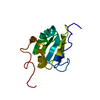
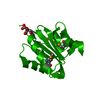
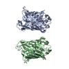
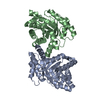
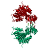
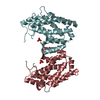
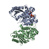
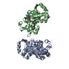
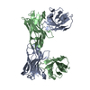
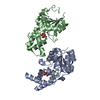
Crystal Structure of sv40 large T antigen origin binding domain with DNA
Components
(21-nt PEN element of the SV40 DNA origin) x 2
large T antigen
Keywords
DNA binding protein/DNA / origin binding domain / protein-DNA complex / replication / DNA binding protein-DNA COMPLEX
Function / homology
Function and homology information
symbiont-mediated suppression of host JAK-STAT cascade via inhibition of JAK1 activity / bidirectional double-stranded viral DNA replication / viral DNA genome replication / symbiont-mediated perturbation of host cell cycle G1/S transition checkpoint / DNA 3'-5' helicase / DNA replication origin binding / 3'-5' DNA helicase activity / single-stranded DNA binding / double-stranded DNA binding / symbiont-mediated perturbation of host ubiquitin-like protein modification ...symbiont-mediated suppression of host JAK-STAT cascade via inhibition of JAK1 activity / bidirectional double-stranded viral DNA replication / viral DNA genome replication / symbiont-mediated perturbation of host cell cycle G1/S transition checkpoint / DNA 3'-5' helicase / DNA replication origin binding / 3'-5' DNA helicase activity / single-stranded DNA binding / double-stranded DNA binding / symbiont-mediated perturbation of host ubiquitin-like protein modification / DNA replication / symbiont-mediated suppression of host innate immune response / symbiont-mediated suppression of host type I interferon-mediated signaling pathway / hydrolase activity / host cell nucleus / ATP hydrolysis activity / DNA-templated transcription / zinc ion binding / ATP binding / identical protein binding Similarity search - Function
Replication Protein E1; Chain: A, - #20 / Replication Protein E1; Chain: A, / Large T antigen, polyomaviridae / Origin of replication binding protein / Large T-antigen (T-ag) origin-binding domain (OBD) profile. / T antigen, Ori-binding / Large T antigen, polyomavirus, C-terminal / Zinc finger, large T-antigen D1-type / Polyomavirus large T antigen C-terminus / Zinc finger large T-antigen (T-ag) D1-type profile. ...Replication Protein E1; Chain: A, - #20 / Replication Protein E1; Chain: A, / Large T antigen, polyomaviridae / Origin of replication binding protein / Large T-antigen (T-ag) origin-binding domain (OBD) profile. / T antigen, Ori-binding / Large T antigen, polyomavirus, C-terminal / Zinc finger, large T-antigen D1-type / Polyomavirus large T antigen C-terminus / Zinc finger large T-antigen (T-ag) D1-type profile. / Zinc finger, large T-antigen D1 domain superfamily / Helicase, superfamily 3, DNA virus / Superfamily 3 helicase of DNA viruses domain profile. / DnaJ molecular chaperone homology domain / dnaJ domain profile. / Chaperone J-domain superfamily / DnaJ domain / P-loop containing nucleoside triphosphate hydrolase / 3-Layer(aba) Sandwich / Alpha Beta Similarity search - Domain/homology
In the structure databanks used in Yorodumi, some data are registered as the other names, "COVID-19 virus" and "2019-nCoV". Here are the details of the virus and the list of structure data.
Jan 31, 2019. EMDB accession codes are about to change! (news from PDBe EMDB page)
EMDB accession codes are about to change! (news from PDBe EMDB page)
The allocation of 4 digits for EMDB accession codes will soon come to an end. Whilst these codes will remain in use, new EMDB accession codes will include an additional digit and will expand incrementally as the available range of codes is exhausted. The current 4-digit format prefixed with “EMD-” (i.e. EMD-XXXX) will advance to a 5-digit format (i.e. EMD-XXXXX), and so on. It is currently estimated that the 4-digit codes will be depleted around Spring 2019, at which point the 5-digit format will come into force.
The EM Navigator/Yorodumi systems omit the EMD- prefix.
Related info.:Q: What is EMD? / ID/Accession-code notation in Yorodumi/EM Navigator
Yorodumi is a browser for structure data from EMDB, PDB, SASBDB, etc.
This page is also the successor to EM Navigator detail page, and also detail information page/front-end page for Omokage search.
The word "yorodu" (or yorozu) is an old Japanese word meaning "ten thousand". "mi" (miru) is to see.
Related info.:EMDB / PDB / SASBDB / Comparison of 3 databanks / Yorodumi Search / Aug 31, 2016. New EM Navigator & Yorodumi / Yorodumi Papers / Jmol/JSmol / Function and homology information / Changes in new EM Navigator and Yorodumi
 Movie
Movie Controller

 Yorodumi
 Open data
 Basic information
 Components
 Keywords
 Function and homology information
 Simian virus 40
 X-RAY DIFFRACTION /
  Authors
 Citation
 Structure visualization
 Downloads & links
 Other downloads
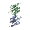


 PDBj
PDBj


 Assembly
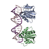

 Mass: 18.015 Da / Num. of mol.: 77 / Source method: isolated from a natural source / Formula: H2O
 Sample preparation
 / Beamline: X29A / Wavelength: 1.1 Å
 Processing