 Movie
Movie Controller

[English] 日本語
 Yorodumi
- PDB-4fbj: Structure of the Cif:Nedd8 complex - Photorhabdus luminescens Cyc... -
+ Open data

- Basic information
| Entry | Database: PDB / ID: 4fbj | ||||||
|---|---|---|---|---|---|---|---|
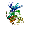
| Title | Structure of the Cif:Nedd8 complex - Photorhabdus luminescens Cycle Inhibiting Factor in complex with human Nedd8 | ||||||
 Components Components |
| ||||||
 Keywords Keywords | CELL CYCLE/PROTEIN BINDING / Effector-Host target complex / Glutamine deamidase / Deamidation / BACTERIAL EFFECTOR / CELL CYCLE-PROTEIN BINDING complex | ||||||
| Function / homology |  Function and homology information Function and homology informationprotein-glutamine glutaminase activity / protein-glutamine glutaminase / symbiont-mediated perturbation of host cell cycle progression / cellular response to camptothecin / protein neddylation / TGF-beta receptor signaling activates SMADs / regulation of proteolysis / regulation of postsynapse assembly / anatomical structure morphogenesis / protein modification process ...protein-glutamine glutaminase activity / protein-glutamine glutaminase / symbiont-mediated perturbation of host cell cycle progression / cellular response to camptothecin / protein neddylation / TGF-beta receptor signaling activates SMADs / regulation of proteolysis / regulation of postsynapse assembly / anatomical structure morphogenesis / protein modification process / Iron uptake and transport / modification-dependent protein catabolic process / protein tag activity / intracellular protein localization / UCH proteinases / Cargo recognition for clathrin-mediated endocytosis / toxin activity / Neddylation / ubiquitin-dependent protein catabolic process / postsynapse / ubiquitin protein ligase binding / regulation of transcription by RNA polymerase II / host cell nucleus / glutamatergic synapse / proteolysis / extracellular exosome / extracellular region / nucleoplasm / nucleus / cytoplasm / cytosol Similarity search - Function | ||||||
| Biological species |  Photorhabdus luminescens subsp. laumondii (bacteria) Photorhabdus luminescens subsp. laumondii (bacteria) Homo sapiens (human) Homo sapiens (human) | ||||||
| Method |  X-RAY DIFFRACTION / SYNCHROTRON / MOLECULAR REPLACEMENT / Resolution: 1.6 Å X-RAY DIFFRACTION / SYNCHROTRON / MOLECULAR REPLACEMENT / Resolution: 1.6 Å | ||||||
 Authors Authors | Crow, A. / Banfield, M.J. | ||||||
 Citation Citation | Journal: Proc.Natl.Acad.Sci.USA / Year: 2012 Title: The molecular basis of Nedd8 deamidation by the bacterial effector protein Cif Authors: Crow, A. / Hughes, R.K. / Taieb, F. / Oswald, E. / Banfield, M.J. | ||||||
| History |
|
- Structure visualization
| Structure viewer | Molecule: MolmilJmol/JSmol |
|---|
- Downloads & links
-Download
| PDBx/mmCIF format | 4fbj.cif.gz | 150.2 KB | Display | PDBx/mmCIF format |
|---|---|---|---|---|
| PDB format | pdb4fbj.ent.gz | 117.8 KB | Display | PDB format |
| PDBx/mmJSON format | 4fbj.json.gz | Tree view | PDBx/mmJSON format | |
| Others |  Other downloads Other downloads |
-Validation report
| Arichive directory | https://data.pdbj.org/pub/pdb/validation_reports/fb/4fbjftp://data.pdbj.org/pub/pdb/validation_reports/fb/4fbj | HTTPS FTP |
|---|
-Related structure data
| Related structure data |  4f8cC 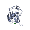 1nddS  3gqjS C: citing same article ( S: Starting model for refinement |
|---|---|
| Similar structure data |


-Links
 PDBj
PDBj

- Assembly
| Deposited unit | 
| ||||||||
|---|---|---|---|---|---|---|---|---|---|
| 1 |
| ||||||||
| Unit cell |
|
-Components
| #1: Protein | Mass: 29637.326 Da / Num. of mol.: 1 / Fragment: Effector domain, UNP residues 53-313 / Mutation: C123S Source method: isolated from a genetically manipulated source Source: (gene. exp.) Photorhabdus luminescens subsp. laumondii (bacteria)Strain: TT01 / Gene: plu2515 / Production host: |
|---|---|
| #2: Protein | Mass: 10044.623 Da / Num. of mol.: 1 Source method: isolated from a genetically manipulated source Source: (gene. exp.) Homo sapiens (human) / Gene: NEDD8 / Production host: |
| #3: Chemical | ChemComp-CL /   Mass: 35.453 Da / Num. of mol.: 1 / Source method: obtained synthetically / Formula: Cl Mass: 35.453 Da / Num. of mol.: 1 / Source method: obtained synthetically / Formula: Cl |
| #4: Water | ChemComp-HOH /  Mass: 18.015 Da / Num. of mol.: 376 / Source method: isolated from a natural source / Formula: H2O Mass: 18.015 Da / Num. of mol.: 376 / Source method: isolated from a natural source / Formula: H2O |
| Nonpolymer details | THE LIGAND IN THE STRUCTURE IS AN UNKNOWN ATOM OR ION WHICH MAY PROBABABLY BE AN CHLORIDE, AND WAS ...THE LIGAND IN THE STRUCTURE IS AN UNKNOWN ATOM OR ION WHICH MAY PROBABABLY |
-Experimental details
-Experiment
| Experiment | Method: X-RAY DIFFRACTION / Number of used crystals: 1 |
|---|
- Sample preparation
| Crystal | Density Matthews: 1.89 Å3/Da / Density % sol: 34.77 % |
|---|---|
| Crystal grow | Temperature: 289 K / Method: vapor diffusion, sitting drop / pH: 6.7 Details: 20% PEG 4000, 200mM sodium acetate, 100mM MES pH 6.7, VAPOR DIFFUSION, SITTING DROP, temperature 289K |
-Data collection
| Diffraction | Mean temperature: 100 K |
|---|---|
| Diffraction source | Source: SYNCHROTRON / Site: Diamond  / Beamline: I02 / Wavelength: 0.9134 Å / Beamline: I02 / Wavelength: 0.9134 Å |
| Detector | Type: ADSC QUANTUM 315r / Detector: CCD / Date: Jan 30, 2011 |
| Radiation | Monochromator: Double crystal monochromator / Protocol: SINGLE WAVELENGTH / Monochromatic (M) / Laue (L): M / Scattering type: x-ray |
| Radiation wavelength | Wavelength: 0.9134 Å / Relative weight: 1 |
| Reflection | Resolution: 1.6→42.6 Å / Num. all: 39093 / Num. obs: 39093 / % possible obs: 96.4 % / Observed criterion σ(F): 1 / Observed criterion σ(I): 1 / Redundancy: 4.4 % / Rsym value: 0.052 / Net I/σ(I): 11.5 |
| Reflection shell | Resolution: 1.6→1.69 Å / Redundancy: 4.3 % / Mean I/σ(I) obs: 3 / Rsym value: 0.288 / % possible all: 96.2 |
- Processing
| Software |
| ||||||||||||||||||||||||||||||||||||||||||||||||||||||||||||||||||||||
|---|---|---|---|---|---|---|---|---|---|---|---|---|---|---|---|---|---|---|---|---|---|---|---|---|---|---|---|---|---|---|---|---|---|---|---|---|---|---|---|---|---|---|---|---|---|---|---|---|---|---|---|---|---|---|---|---|---|---|---|---|---|---|---|---|---|---|---|---|---|---|---|
| Refinement | Method to determine structure: MOLECULAR REPLACEMENT Starting model: PDB ENTRIES 3GQJ and 1NDD Resolution: 1.6→38.2 Å / Cor.coef. Fo:Fc: 0.968 / Cor.coef. Fo:Fc free: 0.939 / SU B: 4.185 / SU ML: 0.078 / Cross valid method: THROUGHOUT / ESU R: 0.146 / ESU R Free: 0.116 / Stereochemistry target values: MAXIMUM LIKELIHOOD / Details: HYDROGENS HAVE BEEN ADDED IN THE RIDING POSITIONS
| ||||||||||||||||||||||||||||||||||||||||||||||||||||||||||||||||||||||
| Solvent computation | Ion probe radii: 0.8 Å / Shrinkage radii: 0.8 Å / VDW probe radii: 1.4 Å / Solvent model: MASK | ||||||||||||||||||||||||||||||||||||||||||||||||||||||||||||||||||||||
| Displacement parameters | Biso mean: 23.474 Å2
| ||||||||||||||||||||||||||||||||||||||||||||||||||||||||||||||||||||||
| Refinement step | Cycle: LAST / Resolution: 1.6→38.2 Å
| ||||||||||||||||||||||||||||||||||||||||||||||||||||||||||||||||||||||
| Refine LS restraints |
| ||||||||||||||||||||||||||||||||||||||||||||||||||||||||||||||||||||||
| LS refinement shell | Resolution: 1.6→1.642 Å / Total num. of bins used: 20
|
