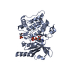
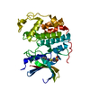
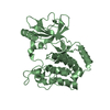
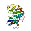
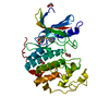
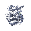
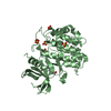
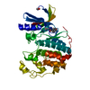
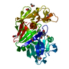
[English] 日本語
Yorodumi
- PDB-4xbu: In vitro Crystal Structure of PAK4 in complex with Inka peptide -

+
Open data


ID or keywords:

Loading...

-
Basic information

Entry
Database: PDB / ID: 4xbu
TitleIn vitro Crystal Structure of PAK4 in complex with Inka peptide
Components
  • Protein FAM212A
  • Serine/threonine-protein kinase PAK 4
KeywordsTransferase/Peptide / PAK4 / Inka / Transferase-Peptide complex
Function / homology
Function and homology information


dendritic spine development / cadherin binding involved in cell-cell adhesion / protein serine/threonine kinase inhibitor activity / Activation of RAC1 / RHOV GTPase cycle / RHOJ GTPase cycle / RHOQ GTPase cycle / regulation of MAPK cascade / CDC42 GTPase cycle / RHOH GTPase cycle ...dendritic spine development / cadherin binding involved in cell-cell adhesion / protein serine/threonine kinase inhibitor activity / Activation of RAC1 / RHOV GTPase cycle / RHOJ GTPase cycle / RHOQ GTPase cycle / regulation of MAPK cascade / CDC42 GTPase cycle / RHOH GTPase cycle / RHOU GTPase cycle / cellular response to organic cyclic compound / RHOG GTPase cycle / RAC2 GTPase cycle / RAC3 GTPase cycle / negative regulation of endothelial cell apoptotic process / cytoskeleton organization / RAC1 GTPase cycle / regulation of cell growth / adherens junction / positive regulation of angiogenesis / cell migration / non-specific serine/threonine protein kinase / protein kinase activity / intracellular signal transduction / cell cycle / phosphorylation / protein serine kinase activity / focal adhesion / protein serine/threonine kinase activity / apoptotic process / protein kinase binding / Golgi apparatus / signal transduction / ATP binding / nucleus / cytoplasm / cytosol
Similarity search - Function
FAM212 domain / PAK4-inhibitor inka / FAM212 family / p21 activated kinase binding domain / CRIB domain superfamily / P21-Rho-binding domain / CRIB domain profile. / P21-Rho-binding domain / CRIB domain / Transferase(Phosphotransferase) domain 1 ...FAM212 domain / PAK4-inhibitor inka / FAM212 family / p21 activated kinase binding domain / CRIB domain superfamily / P21-Rho-binding domain / CRIB domain profile. / P21-Rho-binding domain / CRIB domain / Transferase(Phosphotransferase) domain 1 / Transferase(Phosphotransferase); domain 1 / Phosphorylase Kinase; domain 1 / Phosphorylase Kinase; domain 1 / Protein kinase domain / Protein kinase, ATP binding site / Protein kinases ATP-binding region signature. / Protein kinase domain profile. / Protein kinase domain / Protein kinase-like domain superfamily / 2-Layer Sandwich / Orthogonal Bundle / Mainly Alpha / Alpha Beta
Similarity search - Domain/homology
Serine/threonine-protein kinase PAK 4 / PAK4-inhibitor INKA1
Similarity search - Component
Biological speciesHomo sapiens (human)
MethodX-RAY DIFFRACTION / SYNCHROTRON / MOLECULAR REPLACEMENT / Resolution: 2.06 Å
AuthorsBaskaran, Y. / Ang, K.C. / Anekal, P.V. / Chan, W.L. / Grimes, J.M. / Manser, E. / Robinson, R.C.
Funding support Singapore, 1items
OrganizationGrant numberCountry
Agency for Science, Technology and Research Singapore
CitationJournal: Nat Commun / Year: 2015
Title: An in cellulo-derived structure of PAK4 in complex with its inhibitor Inka1
Authors: Baskaran, Y. / Ang, K.C. / Anekal, P.V. / Chan, W.L. / Grimes, J.M. / Manser, E. / Robinson, R.C.
History
DepositionDec 17, 2014Deposition site: RCSB / Processing site: PDBJ
Revision 1.0Dec 2, 2015Provider: repository / Type: Initial release
Revision 1.1Dec 9, 2015Group: Database references
Revision 1.2Oct 4, 2017Group: Data collection / Derived calculations
Category: diffrn_detector / diffrn_source / pdbx_struct_oper_list
Item: _diffrn_detector.detector / _diffrn_source.pdbx_synchrotron_site / _pdbx_struct_oper_list.symmetry_operation
Revision 1.3Nov 8, 2023Group: Data collection / Database references / Refinement description
Category: chem_comp_atom / chem_comp_bond ...chem_comp_atom / chem_comp_bond / database_2 / pdbx_initial_refinement_model
Item: _database_2.pdbx_DOI / _database_2.pdbx_database_accession

-
Structure visualization

Structure viewerMolecule:
MolmilJmol/JSmol

Downloads & links

-
Assembly

Deposited unit
A: Serine/threonine-protein kinase PAK 4
B: Protein FAM212A


Theoretical massNumber of molelcules
Total (without water)39,7062
Polymers39,7062
Non-polymers00
Water2,702150
1


  • Idetical with deposited unit
  • defined by author&software
TypeNameSymmetry operationNumber
identity operation1_555x,y,z1
Buried area1690 Å2
ΔGint-5 kcal/mol
Surface area14130 Å2
MethodPISA
Unit cell
Length a, b, c (Å)65.620, 65.620, 184.150
Angle α, β, γ (deg.)90.00, 90.00, 90.00
Int Tables number92
Space group name H-MP41212

-
Components

#1: Protein Serine/threonine-protein kinase PAK 4 / p21-activated kinase 4 / PAK-4


Mass: 37046.844 Da / Num. of mol.: 1 / Fragment: UNP residues 286-591
Source method: isolated from a genetically manipulated source
Source: (gene. exp.) Homo sapiens (human) / Gene: PAK4, KIAA1142 / Plasmid: pSY5 / Production host: Escherichia coli (E. coli) / Strain (production host): BL21 Star (DE3) pLysS
References: UniProt: O96013, non-specific serine/threonine protein kinase
#2: Protein/peptide Protein FAM212A


Mass: 2658.968 Da / Num. of mol.: 1 / Fragment: UNP residues 167-190 / Source method: obtained synthetically / Source: (synth.) Homo sapiens (human) / References: UniProt: Q96EL1
#3: Water ChemComp-HOH / water


Mass: 18.015 Da / Num. of mol.: 150 / Source method: isolated from a natural source / Formula: H2O

-
Experimental details

-
Experiment

ExperimentMethod: X-RAY DIFFRACTION

-
Sample preparation

CrystalDensity Matthews: 2.5 Å3/Da / Density % sol: 50.77 % / Description: Bipyramidal
Crystal growTemperature: 298.15 K / Method: vapor diffusion, hanging drop / pH: 8.5 / Details: Tris-HCl, PEG 8000

-
Data collection

DiffractionMean temperature: 80 K
Diffraction sourceSource: SYNCHROTRON / Site: Diamond / Beamline: I24 / Wavelength: 0.9686 Å
DetectorType: DECTRIS PILATUS 6M / Detector: PIXEL / Date: Jul 31, 2014
RadiationProtocol: SINGLE WAVELENGTH / Monochromatic (M) / Laue (L): M / Scattering type: x-ray
Radiation wavelengthWavelength: 0.9686 Å / Relative weight: 1
ReflectionResolution: 2.06→32.81 Å / Num. obs: 25890 / % possible obs: 100 % / Redundancy: 12.8 % / Rmerge(I) obs: 0.074 / Net I/σ(I): 21.2

-
Processing

Software
NameVersionClassification
REFMAC5.8.0049refinement
xia20.3.6.0data reduction
xia20.3.6.0data scaling
xia20.3.6.0data processing
RefinementMethod to determine structure: MOLECULAR REPLACEMENT
Starting model: 4FIE
Resolution: 2.06→32.81 Å / Cor.coef. Fo:Fc: 0.954 / Cor.coef. Fo:Fc free: 0.935 / SU B: 4.589 / SU ML: 0.124 / Cross valid method: THROUGHOUT / ESU R: 0.177 / ESU R Free: 0.165 / Stereochemistry target values: MAXIMUM LIKELIHOOD / Details: HYDROGENS HAVE BEEN ADDED IN THE RIDING POSITIONS
RfactorNum. reflection% reflectionSelection details
Rfree0.24343 1262 4.9 %RANDOM
Rwork0.19856 ---
obs0.20081 24538 99.92 %-
Solvent computationIon probe radii: 0.8 Å / Shrinkage radii: 0.8 Å / VDW probe radii: 1.2 Å / Solvent model: MASK
Displacement parametersBiso mean: 40.805 Å2
Baniso -1Baniso -2Baniso -3
1--0.06 Å2-0 Å20 Å2
2---0.06 Å20 Å2
3---0.12 Å2
Refinement stepCycle: LAST / Resolution: 2.06→32.81 Å
ProteinNucleic acidLigandSolventTotal
Num. atoms2419 0 0 150 2569
Refine LS restraints
Refine-IDTypeDev idealDev ideal targetNumber
X-RAY DIFFRACTIONr_bond_refined_d0.0190.0192467
X-RAY DIFFRACTIONr_bond_other_d0.0010.022456
X-RAY DIFFRACTIONr_angle_refined_deg2.0351.9853340
X-RAY DIFFRACTIONr_angle_other_deg0.97135644
X-RAY DIFFRACTIONr_dihedral_angle_1_deg6.4215304
X-RAY DIFFRACTIONr_dihedral_angle_2_deg35.89923.241108
X-RAY DIFFRACTIONr_dihedral_angle_3_deg15.615445
X-RAY DIFFRACTIONr_dihedral_angle_4_deg16.3011523
X-RAY DIFFRACTIONr_chiral_restr0.1320.2377
X-RAY DIFFRACTIONr_gen_planes_refined0.010.0212738
X-RAY DIFFRACTIONr_gen_planes_other0.0010.02545
X-RAY DIFFRACTIONr_nbd_refined
X-RAY DIFFRACTIONr_nbd_other
X-RAY DIFFRACTIONr_nbtor_refined
X-RAY DIFFRACTIONr_nbtor_other
X-RAY DIFFRACTIONr_xyhbond_nbd_refined
X-RAY DIFFRACTIONr_xyhbond_nbd_other
X-RAY DIFFRACTIONr_metal_ion_refined
X-RAY DIFFRACTIONr_metal_ion_other
X-RAY DIFFRACTIONr_symmetry_vdw_refined
X-RAY DIFFRACTIONr_symmetry_vdw_other
X-RAY DIFFRACTIONr_symmetry_hbond_refined
X-RAY DIFFRACTIONr_symmetry_hbond_other
X-RAY DIFFRACTIONr_symmetry_metal_ion_refined
X-RAY DIFFRACTIONr_symmetry_metal_ion_other
X-RAY DIFFRACTIONr_mcbond_it3.693.7491222
X-RAY DIFFRACTIONr_mcbond_other3.6743.7481221
X-RAY DIFFRACTIONr_mcangle_it4.8325.5981524
X-RAY DIFFRACTIONr_mcangle_other4.835.61525
X-RAY DIFFRACTIONr_scbond_it4.534.2351245
X-RAY DIFFRACTIONr_scbond_other4.5284.2351245
X-RAY DIFFRACTIONr_scangle_it
X-RAY DIFFRACTIONr_scangle_other6.5126.161817
X-RAY DIFFRACTIONr_long_range_B_refined8.12630.7352915
X-RAY DIFFRACTIONr_long_range_B_other8.09130.4712857
X-RAY DIFFRACTIONr_rigid_bond_restr
X-RAY DIFFRACTIONr_sphericity_free
X-RAY DIFFRACTIONr_sphericity_bonded
LS refinement shellResolution: 2.06→2.113 Å / Total num. of bins used: 20
RfactorNum. reflection% reflection
Rfree0.33 90 -
Rwork0.242 1789 -
obs--99.89 %

+
About Yorodumi

-
News

-
Feb 9, 2022. New format data for meta-information of EMDB entries

New format data for meta-information of EMDB entries

  • Version 3 of the EMDB header file is now the official format.
  • The previous official version 1.9 will be removed from the archive.

Related info.:EMDB header

External links:wwPDB to switch to version 3 of the EMDB data model

-
Aug 12, 2020. Covid-19 info

Covid-19 info

URL: https://pdbjlvh1.pdbj.org/emnavi/covid19.php

New page: Covid-19 featured information page in EM Navigator.

Related info.:Covid-19 info / Mar 5, 2020. Novel coronavirus structure data

+
Mar 5, 2020. Novel coronavirus structure data

Novel coronavirus structure data

Related info.:Yorodumi Speices / Aug 12, 2020. Covid-19 info

External links:COVID-19 featured content - PDBj / Molecule of the Month (242):Coronavirus Proteases

+
Jan 31, 2019. EMDB accession codes are about to change! (news from PDBe EMDB page)

EMDB accession codes are about to change! (news from PDBe EMDB page)

  • The allocation of 4 digits for EMDB accession codes will soon come to an end. Whilst these codes will remain in use, new EMDB accession codes will include an additional digit and will expand incrementally as the available range of codes is exhausted. The current 4-digit format prefixed with “EMD-” (i.e. EMD-XXXX) will advance to a 5-digit format (i.e. EMD-XXXXX), and so on. It is currently estimated that the 4-digit codes will be depleted around Spring 2019, at which point the 5-digit format will come into force.
  • The EM Navigator/Yorodumi systems omit the EMD- prefix.

Related info.:Q: What is EMD? / ID/Accession-code notation in Yorodumi/EM Navigator

External links:EMDB Accession Codes are Changing Soon! / Contact to PDBj

+
Jul 12, 2017. Major update of PDB

Major update of PDB

  • wwPDB released updated PDB data conforming to the new PDBx/mmCIF dictionary.
  • This is a major update changing the version number from 4 to 5, and with Remediation, in which all the entries are updated.
  • In this update, many items about electron microscopy experimental information are reorganized (e.g. em_software).
  • Now, EM Navigator and Yorodumi are based on the updated data.

External links:wwPDB Remediation / Enriched Model Files Conforming to OneDep Data Standards Now Available in the PDB FTP Archive

-
Yorodumi

Thousand views of thousand structures

  • Yorodumi is a browser for structure data from EMDB, PDB, SASBDB, etc.
  • This page is also the successor to EM Navigator detail page, and also detail information page/front-end page for Omokage search.
  • The word "yorodu" (or yorozu) is an old Japanese word meaning "ten thousand". "mi" (miru) is to see.

Related info.:EMDB / PDB / SASBDB / Comparison of 3 databanks / Yorodumi Search / Aug 31, 2016. New EM Navigator & Yorodumi / Yorodumi Papers / Jmol/JSmol / Function and homology information / Changes in new EM Navigator and Yorodumi

Read more