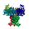
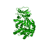
Crystal Structure of Escherichia coli L-arabinose Isomerase (ECAI) complexed with Ribitol
Components
L-arabinose isomerase
Keywords
ISOMERASE / Structural Genomics / PSI-1 / Protein Structure Initiative / New York SGX Research Center for Structural Genomics / NYSGXRC / Sugar Binding
Function / homology
Function and homology information
L-arabinose isomerase / L-arabinose isomerase activity / : / arabinose catabolic process / manganese ion binding / cytosol Similarity search - Function
Resolution: 2.3→20 Å / Cor.coef. Fo:Fc: 0.955 / Cor.coef. Fo:Fc free: 0.935 / SU B: 21.053 / SU ML: 0.218 / Cross valid method: THROUGHOUT / ESU R Free: 0.246 / Stereochemistry target values: MAXIMUM LIKELIHOOD / Details: HYDROGENS HAVE BEEN ADDED IN THE RIDING POSITIONS
Rfactor
Num. reflection
% reflection
Selection details
Rfree
0.25505
3746
5 %
RANDOM
Rwork
0.21184
-
-
-
obs
0.21401
70507
98.31 %
-
Solvent computation
Ion probe radii: 0.8 Å / Shrinkage radii: 0.8 Å / VDW probe radii: 1.2 Å / Solvent model: MASK
Displacement parameters
Biso mean: 28.847 Å2
Baniso -1
Baniso -2
Baniso -3
1-
1.37 Å2
0.68 Å2
0 Å2
2-
-
1.37 Å2
0 Å2
3-
-
-
-2.05 Å2
Refinement step
Cycle: LAST / Resolution: 2.3→20 Å
Protein
Nucleic acid
Ligand
Solvent
Total
Num. atoms
11522
0
37
221
11780
Refine LS restraints
Refine-ID
Type
Dev ideal
Dev ideal target
Number
X-RAY DIFFRACTION
r_bond_refined_d
0.006
0.02
11847
X-RAY DIFFRACTION
r_bond_other_d
0.003
0.02
7718
X-RAY DIFFRACTION
r_angle_refined_deg
1.077
1.945
16096
X-RAY DIFFRACTION
r_angle_other_deg
1.218
3
18794
X-RAY DIFFRACTION
r_dihedral_angle_1_deg
9.187
5
1491
X-RAY DIFFRACTION
r_dihedral_angle_2_deg
36.349
24.378
555
X-RAY DIFFRACTION
r_dihedral_angle_3_deg
15.9
15
1714
X-RAY DIFFRACTION
r_dihedral_angle_4_deg
18.233
15
59
X-RAY DIFFRACTION
r_chiral_restr
0.062
0.2
1773
X-RAY DIFFRACTION
r_gen_planes_refined
0.004
0.021
13420
X-RAY DIFFRACTION
r_gen_planes_other
0.002
0.02
2459
X-RAY DIFFRACTION
r_rigid_bond_restr
8.025
3
19565
X-RAY DIFFRACTION
r_sphericity_free
42.407
5
59
X-RAY DIFFRACTION
r_sphericity_bonded
7.585
5
19457
LS refinement shell
Resolution: 2.3→2.36 Å / Total num. of bins used: 20
Rfactor
Num. reflection
% reflection
Rfree
0.371
225
-
Rwork
0.308
4068
-
obs
-
-
83.9 %
Refinement TLS params.
Method: refined / Refine-ID: X-RAY DIFFRACTION
ID
L11 (°2)
L12 (°2)
L13 (°2)
L22 (°2)
L23 (°2)
L33 (°2)
S11 (Å °)
S12 (Å °)
S13 (Å °)
S21 (Å °)
S22 (Å °)
S23 (Å °)
S31 (Å °)
S32 (Å °)
S33 (Å °)
T11 (Å2)
T12 (Å2)
T13 (Å2)
T22 (Å2)
T23 (Å2)
T33 (Å2)
Origin x (Å)
Origin y (Å)
Origin z (Å)
1
0.6913
-0.0254
-0.0101
0.223
-0.2567
1.4406
-0.0579
0.0952
0.008
-0.174
0.0113
0.0646
-0.012
0.2544
0.0466
0.4644
-0.1032
-0.0378
0.0831
0.0371
0.2479
61.803
4.532
2.95
2
0.2411
-0.0799
0.2296
0.4397
-0.3196
1.6565
-0.0119
0.0239
-0.1157
-0.0409
0.0971
0.1792
0.4535
-0.2244
-0.0852
0.4903
-0.0952
-0.0159
0.0596
0.0382
0.3707
39.116
-23.325
34.661
3
0.2647
0.2412
0.1308
1.0041
-0.5396
1.5405
0.0378
0.134
-0.1707
-0.2447
-0.2547
-0.1301
0.5115
1.0044
0.2168
0.5309
0.4016
0.0845
0.6853
0.1216
0.2279
85.849
-23.309
34.341
Refinement TLS group
ID
Refine-ID
Refine TLS-ID
Auth asym-ID
Auth seq-ID
1
X-RAY DIFFRACTION
1
A
1 - 174
2
X-RAY DIFFRACTION
1
A
175 - 354
3
X-RAY DIFFRACTION
1
A
355 - 498
4
X-RAY DIFFRACTION
2
B
1 - 174
5
X-RAY DIFFRACTION
2
B
175 - 354
6
X-RAY DIFFRACTION
2
B
355 - 498
7
X-RAY DIFFRACTION
3
C
1 - 174
8
X-RAY DIFFRACTION
3
C
175 - 354
9
X-RAY DIFFRACTION
3
C
355 - 498
+
About Yorodumi
-
News
-
Feb 9, 2022. New format data for meta-information of EMDB entries
New format data for meta-information of EMDB entries
Version 3 of the EMDB header file is now the official format.
The previous official version 1.9 will be removed from the archive.
In the structure databanks used in Yorodumi, some data are registered as the other names, "COVID-19 virus" and "2019-nCoV". Here are the details of the virus and the list of structure data.
Jan 31, 2019. EMDB accession codes are about to change! (news from PDBe EMDB page)
EMDB accession codes are about to change! (news from PDBe EMDB page)
The allocation of 4 digits for EMDB accession codes will soon come to an end. Whilst these codes will remain in use, new EMDB accession codes will include an additional digit and will expand incrementally as the available range of codes is exhausted. The current 4-digit format prefixed with “EMD-” (i.e. EMD-XXXX) will advance to a 5-digit format (i.e. EMD-XXXXX), and so on. It is currently estimated that the 4-digit codes will be depleted around Spring 2019, at which point the 5-digit format will come into force.
The EM Navigator/Yorodumi systems omit the EMD- prefix.
Related info.:Q: What is EMD? / ID/Accession-code notation in Yorodumi/EM Navigator
Yorodumi is a browser for structure data from EMDB, PDB, SASBDB, etc.
This page is also the successor to EM Navigator detail page, and also detail information page/front-end page for Omokage search.
The word "yorodu" (or yorozu) is an old Japanese word meaning "ten thousand". "mi" (miru) is to see.
Related info.:EMDB / PDB / SASBDB / Comparison of 3 databanks / Yorodumi Search / Aug 31, 2016. New EM Navigator & Yorodumi / Yorodumi Papers / Jmol/JSmol / Function and homology information / Changes in new EM Navigator and Yorodumi
 Movie
Movie Controller

 Yorodumi
 Open data
 Basic information
 Components
 Keywords
 Function and homology information
 X-RAY DIFFRACTION /
  Authors
 Citation
 Structure visualization
 Downloads & links
 Other downloads


 PDBj
PDBj

 Assembly
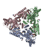

 Mass: 54.938 Da / Num. of mol.: 3 / Source method: obtained synthetically / Formula: Mn
 Mass: 152.146 Da / Num. of mol.: 3
 Mass: 60.052 Da / Num. of mol.: 1 / Source method: obtained synthetically / Formula: C2H4O2
 Mass: 18.015 Da / Num. of mol.: 221 / Source method: isolated from a natural source / Formula: H2O
 Sample preparation
 Processing