 Movie
Movie Controller

[English] 日本語
 Yorodumi
- PDB-4lql: Crystal structure of L-arabinose isomerase from Lactobacillus fer... -
+ Open data

- Basic information
| Entry | Database: PDB / ID: 4lql | ||||||
|---|---|---|---|---|---|---|---|
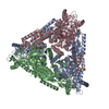
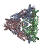
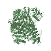
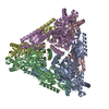
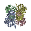
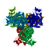
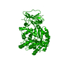
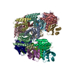
| Title | Crystal structure of L-arabinose isomerase from Lactobacillus fermentum CGMCC2921 | ||||||
 Components Components | L-arabinose isomerase | ||||||
 Keywords Keywords | ISOMERASE / hexamer / isomerization | ||||||
| Function / homology |  Function and homology information Function and homology informationL-arabinose isomerase / L-arabinose isomerase activity / : / metal ion binding / cytosol Similarity search - Function | ||||||
| Biological species |  Lactobacillus fermentum (bacteria) Lactobacillus fermentum (bacteria) | ||||||
| Method |  X-RAY DIFFRACTION / SYNCHROTRON / MOLECULAR REPLACEMENT / Resolution: 3.232 Å X-RAY DIFFRACTION / SYNCHROTRON / MOLECULAR REPLACEMENT / Resolution: 3.232 Å | ||||||
 Authors Authors | Xu, Z. | ||||||
 Citation Citation | Journal: to be published Title: Crystal structure of L-arabinose isomerase from Lactobacillus fermentum CGMCC2921 Authors: Xu, Z. | ||||||
| History |
|
- Structure visualization
| Structure viewer | Molecule: MolmilJmol/JSmol |
|---|
- Downloads & links
-Download
| PDBx/mmCIF format | 4lql.cif.gz | 492.9 KB | Display | PDBx/mmCIF format |
|---|---|---|---|---|
| PDB format | pdb4lql.ent.gz | 382.5 KB | Display | PDB format |
| PDBx/mmJSON format | 4lql.json.gz | Tree view | PDBx/mmJSON format | |
| Others |  Other downloads Other downloads |
-Validation report
| Arichive directory | https://data.pdbj.org/pub/pdb/validation_reports/lq/4lqlftp://data.pdbj.org/pub/pdb/validation_reports/lq/4lql | HTTPS FTP |
|---|
-Related structure data
| Related structure data | 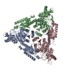 2ajtS S: Starting model for refinement |
|---|---|
| Similar structure data |
-Links
 PDBj
PDBj- Assembly
| Deposited unit | 
| ||||||||
|---|---|---|---|---|---|---|---|---|---|
| 1 |
| ||||||||
| Unit cell |
|
-Components
| #1: Protein | Mass: 53492.164 Da / Num. of mol.: 6 / Source method: isolated from a natural source / Source: (natural) Lactobacillus fermentum (bacteria) / References: UniProt: D9ILD9, L-arabinose isomerase |
|---|
-Experimental details
-Experiment
| Experiment | Method: X-RAY DIFFRACTION / Number of used crystals: 1 |
|---|
- Sample preparation
| Crystal | Density Matthews: 2.29 Å3/Da / Density % sol: 46.19 % |
|---|---|
| Crystal grow | Temperature: 293 K / Method: vapor diffusion, sitting drop / pH: 6 Details: 0.1M Bis-Tris, 25% PEGMME5000, pH 6.0, VAPOR DIFFUSION, SITTING DROP, temperature 293K |
-Data collection
| Diffraction | Mean temperature: 173 K |
|---|---|
| Diffraction source | Source: SYNCHROTRON / Site: SSRF  / Beamline: BL17U / Wavelength: 1.054 Å / Beamline: BL17U / Wavelength: 1.054 Å |
| Detector | Type: ADSC QUANTUM 315r / Detector: CCD / Date: May 30, 2012 |
| Radiation | Protocol: SINGLE WAVELENGTH / Monochromatic (M) / Laue (L): M / Scattering type: x-ray |
| Radiation wavelength | Wavelength: 1.054 Å / Relative weight: 1 |
| Reflection | Resolution: 3.23→46.59 Å / Num. all: 90230 / Num. obs: 47550 / % possible obs: 99 % / Biso Wilson estimate: 94.79 Å2 / Rmerge(I) obs: 0.102 |
| Reflection shell | Highest resolution: 3.23 Å / % possible all: 99 |
- Processing
| Software |
| ||||||||||||||||||||||||||||||||||||||||||||||||||||||||||||||||||||||||||||||||||||||||||||||||||||||||||||||||||||||||||||||
|---|---|---|---|---|---|---|---|---|---|---|---|---|---|---|---|---|---|---|---|---|---|---|---|---|---|---|---|---|---|---|---|---|---|---|---|---|---|---|---|---|---|---|---|---|---|---|---|---|---|---|---|---|---|---|---|---|---|---|---|---|---|---|---|---|---|---|---|---|---|---|---|---|---|---|---|---|---|---|---|---|---|---|---|---|---|---|---|---|---|---|---|---|---|---|---|---|---|---|---|---|---|---|---|---|---|---|---|---|---|---|---|---|---|---|---|---|---|---|---|---|---|---|---|---|---|---|---|
| Refinement | Method to determine structure: MOLECULAR REPLACEMENT Starting model: 2AJT Resolution: 3.232→44.849 Å / Occupancy max: 1 / Occupancy min: 1 / FOM work R set: 0.5237 / SU ML: 0.85 / σ(F): 1.35 / Phase error: 50.03 / Stereochemistry target values: ML
| ||||||||||||||||||||||||||||||||||||||||||||||||||||||||||||||||||||||||||||||||||||||||||||||||||||||||||||||||||||||||||||||
| Solvent computation | Shrinkage radii: 0.9 Å / VDW probe radii: 1.11 Å / Solvent model: FLAT BULK SOLVENT MODEL | ||||||||||||||||||||||||||||||||||||||||||||||||||||||||||||||||||||||||||||||||||||||||||||||||||||||||||||||||||||||||||||||
| Displacement parameters | Biso max: 265.02 Å2 / Biso mean: 80.7835 Å2 / Biso min: 13.76 Å2 | ||||||||||||||||||||||||||||||||||||||||||||||||||||||||||||||||||||||||||||||||||||||||||||||||||||||||||||||||||||||||||||||
| Refinement step | Cycle: LAST / Resolution: 3.232→44.849 Å
| ||||||||||||||||||||||||||||||||||||||||||||||||||||||||||||||||||||||||||||||||||||||||||||||||||||||||||||||||||||||||||||||
| Refine LS restraints |
| ||||||||||||||||||||||||||||||||||||||||||||||||||||||||||||||||||||||||||||||||||||||||||||||||||||||||||||||||||||||||||||||
| LS refinement shell | Refine-ID: X-RAY DIFFRACTION / Total num. of bins used: 17
|
