 Movie
Movie Controller

[English] 日本語
 Yorodumi
- PDB-1k70: The Structure of Escherichia coli Cytosine Deaminase bound to 4-H... -
+ Open data

- Basic information
| Entry | Database: PDB / ID: 1k70 | |||||||||
|---|---|---|---|---|---|---|---|---|---|---|
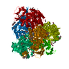
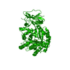
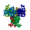
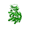
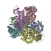
| Title | The Structure of Escherichia coli Cytosine Deaminase bound to 4-Hydroxy-3,4-Dihydro-1H-Pyrimidin-2-one | |||||||||
 Components Components | Cytosine Deaminase | |||||||||
 Keywords Keywords | HYDROLASE / cytosine deaminase / alpha-beta barrel / hexamer / conformational change | |||||||||
| Function / homology |  Function and homology information Function and homology informationcytosine catabolic process / isoguanine deaminase activity / cytosine deaminase / cytosine deaminase activity / Hydrolases; Acting on carbon-nitrogen bonds, other than peptide bonds; In cyclic amidines / ferrous iron binding / zinc ion binding / identical protein binding / cytosol Similarity search - Function | |||||||||
| Biological species |  | |||||||||
| Method |  X-RAY DIFFRACTION / MAD / Resolution: 1.8 Å X-RAY DIFFRACTION / MAD / Resolution: 1.8 Å | |||||||||
 Authors Authors | Ireton, G.C. / McDermott, G. / Black, M.E. / Stoddard, B.L. | |||||||||
 Citation Citation | Journal: J.Mol.Biol. / Year: 2002 Title: The structure of Escherichia coli cytosine deaminase. Authors: Ireton, G.C. / McDermott, G. / Black, M.E. / Stoddard, B.L. | |||||||||
| History |
|
- Structure visualization
| Structure viewer | Molecule: MolmilJmol/JSmol |
|---|
- Downloads & links
-Download
| PDBx/mmCIF format | 1k70.cif.gz | 103.4 KB | Display | PDBx/mmCIF format |
|---|---|---|---|---|
| PDB format | pdb1k70.ent.gz | 79 KB | Display | PDB format |
| PDBx/mmJSON format | 1k70.json.gz | Tree view | PDBx/mmJSON format | |
| Others |  Other downloads Other downloads |
-Validation report
| Arichive directory | https://data.pdbj.org/pub/pdb/validation_reports/k7/1k70ftp://data.pdbj.org/pub/pdb/validation_reports/k7/1k70 | HTTPS FTP |
|---|
-Related structure data


-Links
 PDBj
PDBj

- Assembly
| Deposited unit | 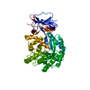
| ||||||||
|---|---|---|---|---|---|---|---|---|---|
| 1 | x 6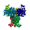
| ||||||||
| Unit cell |
| ||||||||
| Components on special symmetry positions |
| ||||||||
| Details | he biological assembly is a hexamer of three domain swapped dimers generated form the monomer in the asymmetric unit by the operations:-y,x-y,z;y-x,-x,z; y,x,-z;x-y,-y,-z and -x,y-x,z. |
-Components
| #1: Protein | Mass: 47513.594 Da / Num. of mol.: 1 Source method: isolated from a genetically manipulated source Source: (gene. exp.) |
|---|---|
| #2: Chemical | ChemComp-FE /   Mass: 55.845 Da / Num. of mol.: 1 / Source method: obtained synthetically / Formula: Fe Mass: 55.845 Da / Num. of mol.: 1 / Source method: obtained synthetically / Formula: Fe |
| #3: Chemical | ChemComp-HPY /   Mass: 114.103 Da / Num. of mol.: 1 / Source method: obtained synthetically / Formula: C4H6N2O2 Mass: 114.103 Da / Num. of mol.: 1 / Source method: obtained synthetically / Formula: C4H6N2O2 |
| #4: Water | ChemComp-HOH /  Mass: 18.015 Da / Num. of mol.: 367 / Source method: isolated from a natural source / Formula: H2O Mass: 18.015 Da / Num. of mol.: 367 / Source method: isolated from a natural source / Formula: H2O |
-Experimental details
-Experiment
| Experiment | Method: X-RAY DIFFRACTION / Number of used crystals: 1 |
|---|
- Sample preparation
| Crystal | Density Matthews: 2.92 Å3/Da / Density % sol: 57.84 % | |||||||||||||||||||||||||||||||||||
|---|---|---|---|---|---|---|---|---|---|---|---|---|---|---|---|---|---|---|---|---|---|---|---|---|---|---|---|---|---|---|---|---|---|---|---|---|
| Crystal grow | Temperature: 298 K / Method: vapor diffusion, hanging drop / pH: 7.5 Details: PEG 8000, magnesium chloride, Hepes, pH 7.5, VAPOR DIFFUSION, HANGING DROP, temperature 298K | |||||||||||||||||||||||||||||||||||
| Crystal grow | *PLUS PH range low: 7.7 / PH range high: 7.3 | |||||||||||||||||||||||||||||||||||
| Components of the solutions | *PLUS
|
-Data collection
| Diffraction | Mean temperature: 100 K |
|---|---|
| Diffraction source | Source: ROTATING ANODE / Type: RIGAKU / Wavelength: 1.5418 |
| Detector | Type: RIGAKU RAXIS IV / Detector: IMAGE PLATE / Date: Apr 8, 2001 |
| Radiation | Monochromator: Yale Mirrors / Protocol: SINGLE WAVELENGTH / Monochromatic (M) / Laue (L): M / Scattering type: x-ray |
| Radiation wavelength | Wavelength: 1.5418 Å / Relative weight: 1 |
| Reflection | Resolution: 1.8→20 Å / Num. all: 50003 / Num. obs: 50003 / % possible obs: 99 % / Observed criterion σ(F): 0 / Observed criterion σ(I): 0 / Biso Wilson estimate: 18 Å2 / Rmerge(I) obs: 0.047 |
| Reflection shell | Resolution: 1.8→1.86 Å / Rmerge(I) obs: 0.174 / Mean I/σ(I) obs: 12.7 / % possible all: 92.2 |
| Reflection | *PLUS Highest resolution: 1.8 Å / Lowest resolution: 30 Å / Num. obs: 51159 / Num. measured all: 331182 / Rmerge(I) obs: 0.048 |
| Reflection shell | *PLUS % possible obs: 92.2 % / Mean I/σ(I) obs: 12.6 |
- Processing
| Software |
| |||||||||||||||||||||||||
|---|---|---|---|---|---|---|---|---|---|---|---|---|---|---|---|---|---|---|---|---|---|---|---|---|---|---|
| Refinement | Method to determine structure: MAD / Resolution: 1.8→19.97 Å / Rfactor Rfree error: 0.004 / Data cutoff high absF: 710238.17 / Data cutoff low absF: 0 / Isotropic thermal model: RESTRAINED / Cross valid method: THROUGHOUT / σ(F): 0 / Stereochemistry target values: Engh & Huber
| |||||||||||||||||||||||||
| Solvent computation | Solvent model: FLAT MODEL / Bsol: 57.0446 Å2 / ksol: 0.402847 e/Å3 | |||||||||||||||||||||||||
| Displacement parameters | Biso mean: 15.9 Å2
| |||||||||||||||||||||||||
| Refine analyze |
| |||||||||||||||||||||||||
| Refinement step | Cycle: LAST / Resolution: 1.8→19.97 Å
| |||||||||||||||||||||||||
| Refine LS restraints |
| |||||||||||||||||||||||||
| LS refinement shell | Resolution: 1.8→1.91 Å / Rfactor Rfree error: 0.015 / Total num. of bins used: 6
| |||||||||||||||||||||||||
| Xplor file |
| |||||||||||||||||||||||||
| Software | *PLUS Name: CNS / Version: 1 / Classification: refinement | |||||||||||||||||||||||||
| Refinement | *PLUS Lowest resolution: 20 Å / σ(F): 0 / % reflection Rfree: 5 % / Rfactor obs: 0.177 | |||||||||||||||||||||||||
| Solvent computation | *PLUS | |||||||||||||||||||||||||
| Displacement parameters | *PLUS Biso mean: 15.9 Å2 | |||||||||||||||||||||||||
| Refine LS restraints | *PLUS
| |||||||||||||||||||||||||
| LS refinement shell | *PLUS Rfactor Rfree: 0.295 / % reflection Rfree: 5.1 % / Rfactor Rwork: 0.273 |
