 Movie
Movie Controller

[English] 日本語
 Yorodumi
- PDB-4r85: Crystal structure of 5-methylcytosine deaminase from Klebsiella p... -
+ Open data

- Basic information
| Entry | Database: PDB / ID: 4r85 | |||||||||
|---|---|---|---|---|---|---|---|---|---|---|
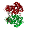
| Title | Crystal structure of 5-methylcytosine deaminase from Klebsiella pneumoniae liganded with 5-methylcytosine | |||||||||
 Components Components | Cytosine deaminase | |||||||||
 Keywords Keywords | HYDROLASE / Amidohydrolase fold / 5-methylcytosine deaminase / 5-methylcytosine | |||||||||
| Function / homology |  Function and homology information Function and homology information: / cytosine catabolic process / isoguanine deaminase activity / cytosine deaminase / cytosine deaminase activity / metal ion binding Similarity search - Function | |||||||||
| Biological species |  Klebsiella pneumoniae (bacteria) Klebsiella pneumoniae (bacteria) | |||||||||
| Method |  X-RAY DIFFRACTION / SYNCHROTRON / MOLECULAR REPLACEMENT / Resolution: 1.802 Å X-RAY DIFFRACTION / SYNCHROTRON / MOLECULAR REPLACEMENT / Resolution: 1.802 Å | |||||||||
 Authors Authors | Fedorov, A.A. / Fedorov, E.V. / Hitchcock, D.S. / Raushel, F.M. / Almo, S.C. | |||||||||
 Citation Citation | Journal: To be Published Title: Crystal structure of 5-methylcytosine deaminase from Klebsiella pneumoniae liganded with 5-methylcytosine Authors: Fedorov, A.A. / Fedorov, E.V. / Hitchcock, D.S. / Raushel, F.M. / Almo, S.C. | |||||||||
| History |
|
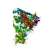
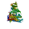
- Structure visualization
| Structure viewer | Molecule: MolmilJmol/JSmol |
|---|
- Downloads & links
-Download
| PDBx/mmCIF format | 4r85.cif.gz | 1016.5 KB | Display | PDBx/mmCIF format |
|---|---|---|---|---|
| PDB format | pdb4r85.ent.gz | 848.1 KB | Display | PDB format |
| PDBx/mmJSON format | 4r85.json.gz | Tree view | PDBx/mmJSON format | |
| Others |  Other downloads Other downloads |
-Validation report
| Arichive directory | https://data.pdbj.org/pub/pdb/validation_reports/r8/4r85ftp://data.pdbj.org/pub/pdb/validation_reports/r8/4r85 | HTTPS FTP |
|---|
-Related structure data
| Related structure data |  4jnp S: Starting model for refinement |
|---|---|
| Similar structure data |


-Links
 PDBj
PDBj


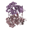
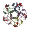
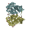
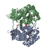
- Assembly
| Deposited unit | 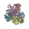
| ||||||||
|---|---|---|---|---|---|---|---|---|---|
| 1 | 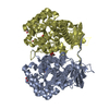
| ||||||||
| 2 | 
| ||||||||
| 3 | 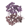
| ||||||||
| 4 |
| ||||||||
| Unit cell |
|
-Components
| #1: Protein | Mass: 48067.453 Da / Num. of mol.: 6 Source method: isolated from a genetically manipulated source Source: (gene. exp.) Klebsiella pneumoniae (bacteria) / Strain: 30660/NJST258_1 / Gene: KPNJ1_03949 / Production host: References: UniProt: W8V4R8, UniProt: A0A0E1CHI1*PLUS, cytosine deaminase #2: Chemical | ChemComp-17E /   Mass: 125.129 Da / Num. of mol.: 6 / Source method: obtained synthetically / Formula: C5H7N3O Mass: 125.129 Da / Num. of mol.: 6 / Source method: obtained synthetically / Formula: C5H7N3O#3: Chemical | ChemComp-FE2 /   Mass: 55.845 Da / Num. of mol.: 6 / Source method: obtained synthetically / Formula: Fe Mass: 55.845 Da / Num. of mol.: 6 / Source method: obtained synthetically / Formula: Fe#4: Chemical | ChemComp-GOL /   Mass: 92.094 Da / Num. of mol.: 18 / Source method: obtained synthetically / Formula: C3H8O3 Mass: 92.094 Da / Num. of mol.: 18 / Source method: obtained synthetically / Formula: C3H8O3#5: Water | ChemComp-HOH / |  Mass: 18.015 Da / Num. of mol.: 1714 / Source method: isolated from a natural source / Formula: H2O Mass: 18.015 Da / Num. of mol.: 1714 / Source method: isolated from a natural source / Formula: H2O |
|---|
-Experimental details
-Experiment
| Experiment | Method: X-RAY DIFFRACTION / Number of used crystals: 1 |
|---|
- Sample preparation
| Crystal | Density Matthews: 2.63 Å3/Da / Density % sol: 53.28 % |
|---|---|
| Crystal grow | Temperature: 293 K / Method: vapor diffusion, sitting drop / pH: 7 Details: 20% PEG 3350, 0.15M DL-MALIC ACID, pH 7.0, VAPOR DIFFUSION, SITTING DROP, temperature 293.0K |
-Data collection
| Diffraction | Mean temperature: 100 K |
|---|---|
| Diffraction source | Source: SYNCHROTRON / Site: NSLS  / Beamline: X29A / Wavelength: 1.075 Å / Beamline: X29A / Wavelength: 1.075 Å |
| Detector | Type: ADSC QUANTUM 315r / Detector: CCD / Date: Mar 10, 2013 |
| Radiation | Monochromator: SI 111 CHANNEL / Protocol: SINGLE WAVELENGTH / Monochromatic (M) / Laue (L): M / Scattering type: x-ray |
| Radiation wavelength | Wavelength: 1.075 Å / Relative weight: 1 |
| Reflection | Resolution: 1.802→40.174 Å / Num. obs: 265794 / % possible obs: 96.8 % / Observed criterion σ(F): 0 / Observed criterion σ(I): 0 |
- Processing
| Software |
| |||||||||||||||||||||||||||||||||||||||||||||||||||||||||||||||||||||||||||||||||||||||||||||||||||||||||||||||||||||||||||||||||||||||||||||||||||||||||||||||||||||||||||||||||||||||||||||||||||||||||||||||||||||||||
|---|---|---|---|---|---|---|---|---|---|---|---|---|---|---|---|---|---|---|---|---|---|---|---|---|---|---|---|---|---|---|---|---|---|---|---|---|---|---|---|---|---|---|---|---|---|---|---|---|---|---|---|---|---|---|---|---|---|---|---|---|---|---|---|---|---|---|---|---|---|---|---|---|---|---|---|---|---|---|---|---|---|---|---|---|---|---|---|---|---|---|---|---|---|---|---|---|---|---|---|---|---|---|---|---|---|---|---|---|---|---|---|---|---|---|---|---|---|---|---|---|---|---|---|---|---|---|---|---|---|---|---|---|---|---|---|---|---|---|---|---|---|---|---|---|---|---|---|---|---|---|---|---|---|---|---|---|---|---|---|---|---|---|---|---|---|---|---|---|---|---|---|---|---|---|---|---|---|---|---|---|---|---|---|---|---|---|---|---|---|---|---|---|---|---|---|---|---|---|---|---|---|---|---|---|---|---|---|---|---|---|---|---|---|---|---|---|---|---|
| Refinement | Method to determine structure: MOLECULAR REPLACEMENT Starting model: PDB entry 4JNP 4jnp Resolution: 1.802→40.174 Å / SU ML: 0.18 / σ(F): 0 / Phase error: 20.19 / Stereochemistry target values: ML
| |||||||||||||||||||||||||||||||||||||||||||||||||||||||||||||||||||||||||||||||||||||||||||||||||||||||||||||||||||||||||||||||||||||||||||||||||||||||||||||||||||||||||||||||||||||||||||||||||||||||||||||||||||||||||
| Solvent computation | Shrinkage radii: 0.9 Å / VDW probe radii: 1.11 Å / Solvent model: FLAT BULK SOLVENT MODEL | |||||||||||||||||||||||||||||||||||||||||||||||||||||||||||||||||||||||||||||||||||||||||||||||||||||||||||||||||||||||||||||||||||||||||||||||||||||||||||||||||||||||||||||||||||||||||||||||||||||||||||||||||||||||||
| Refinement step | Cycle: LAST / Resolution: 1.802→40.174 Å
| |||||||||||||||||||||||||||||||||||||||||||||||||||||||||||||||||||||||||||||||||||||||||||||||||||||||||||||||||||||||||||||||||||||||||||||||||||||||||||||||||||||||||||||||||||||||||||||||||||||||||||||||||||||||||
| Refine LS restraints |
| |||||||||||||||||||||||||||||||||||||||||||||||||||||||||||||||||||||||||||||||||||||||||||||||||||||||||||||||||||||||||||||||||||||||||||||||||||||||||||||||||||||||||||||||||||||||||||||||||||||||||||||||||||||||||
| LS refinement shell |
| |||||||||||||||||||||||||||||||||||||||||||||||||||||||||||||||||||||||||||||||||||||||||||||||||||||||||||||||||||||||||||||||||||||||||||||||||||||||||||||||||||||||||||||||||||||||||||||||||||||||||||||||||||||||||
| Refinement TLS params. | Method: refined / Refine-ID: X-RAY DIFFRACTION
| |||||||||||||||||||||||||||||||||||||||||||||||||||||||||||||||||||||||||||||||||||||||||||||||||||||||||||||||||||||||||||||||||||||||||||||||||||||||||||||||||||||||||||||||||||||||||||||||||||||||||||||||||||||||||
| Refinement TLS group |
|
