 Movie
Movie Controller

[English] 日本語
 Yorodumi
- PDB-4ew1: High resolution structure of human glycinamide ribonucleotide tra... -
+ Open data

- Basic information
| Entry | Database: PDB / ID: 4ew1 | ||||||
|---|---|---|---|---|---|---|---|
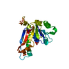
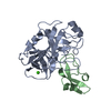
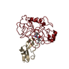
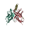
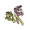
| Title | High resolution structure of human glycinamide ribonucleotide transformylase in apo form. | ||||||
 Components Components | Trifunctional purine biosynthetic protein adenosine-3 | ||||||
 Keywords Keywords | TRANSFERASE | ||||||
| Function / homology |  Function and homology information Function and homology informationphosphoribosylamine-glycine ligase / phosphoribosylglycinamide formyltransferase 1 / adenine biosynthetic process / phosphoribosylformylglycinamidine cyclo-ligase / phosphoribosylformylglycinamidine cyclo-ligase activity / phosphoribosylglycinamide formyltransferase activity / purine ribonucleoside monophosphate biosynthetic process / phosphoribosylamine-glycine ligase activity / brainstem development / 'de novo' XMP biosynthetic process ...phosphoribosylamine-glycine ligase / phosphoribosylglycinamide formyltransferase 1 / adenine biosynthetic process / phosphoribosylformylglycinamidine cyclo-ligase / phosphoribosylformylglycinamidine cyclo-ligase activity / phosphoribosylglycinamide formyltransferase activity / purine ribonucleoside monophosphate biosynthetic process / phosphoribosylamine-glycine ligase activity / brainstem development / 'de novo' XMP biosynthetic process / Purine ribonucleoside monophosphate biosynthesis / 'de novo' AMP biosynthetic process / purine nucleotide biosynthetic process / GMP biosynthetic process / 'de novo' IMP biosynthetic process / cerebellum development / cerebral cortex development / extracellular exosome / ATP binding / metal ion binding / cytosol Similarity search - Function | ||||||
| Biological species |  Homo sapiens (human) Homo sapiens (human) | ||||||
| Method |  X-RAY DIFFRACTION / SYNCHROTRON / MOLECULAR REPLACEMENT / Resolution: 1.522 Å X-RAY DIFFRACTION / SYNCHROTRON / MOLECULAR REPLACEMENT / Resolution: 1.522 Å | ||||||
 Authors Authors | Connelly, S. / DeMartino, K. / Boger, D.L. / Wilson, I.A. | ||||||
 Citation Citation | Journal: Biochemistry / Year: 2013 Title: Biological and Structural Evaluation of 10R- and 10S-Methylthio-DDACTHF Reveals a New Role for Sulfur in Inhibition of Glycinamide Ribonucleotide Transformylase. Authors: Connelly, S. / Demartino, J.K. / Boger, D.L. / Wilson, I.A. | ||||||
| History |
|
- Structure visualization
| Structure viewer | Molecule: MolmilJmol/JSmol |
|---|
- Downloads & links
-Download
| PDBx/mmCIF format | 4ew1.cif.gz | 107.8 KB | Display | PDBx/mmCIF format |
|---|---|---|---|---|
| PDB format | pdb4ew1.ent.gz | 83.3 KB | Display | PDB format |
| PDBx/mmJSON format | 4ew1.json.gz | Tree view | PDBx/mmJSON format | |
| Others |  Other downloads Other downloads |
-Validation report
| Arichive directory | https://data.pdbj.org/pub/pdb/validation_reports/ew/4ew1ftp://data.pdbj.org/pub/pdb/validation_reports/ew/4ew1 | HTTPS FTP |
|---|
-Related structure data
| Related structure data |  4ew2C  4ew3C  1meoS S: Starting model for refinement C: citing same article ( |
|---|---|
| Similar structure data |


-Links
 PDBj
PDBj


- Assembly
| Deposited unit | 
| |||||||||
|---|---|---|---|---|---|---|---|---|---|---|
| 1 |
| |||||||||
| 2 | 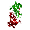
| |||||||||
| Unit cell |
| |||||||||
| Components on special symmetry positions |
|
-Components
| #1: Protein | Mass: 22678.941 Da / Num. of mol.: 1 / Fragment: Residues 808-1010 Source method: isolated from a genetically manipulated source Source: (gene. exp.) Homo sapiens (human) / Gene: GART, PGFT, PRGS / Plasmid: pet22b / Production host:  References: UniProt: P22102, phosphoribosylamine-glycine ligase, phosphoribosylformylglycinamidine cyclo-ligase, phosphoribosylglycinamide formyltransferase 1 | ||
|---|---|---|---|
| #2: Chemical | ChemComp-PO4 /   Mass: 94.971 Da / Num. of mol.: 1 / Source method: obtained synthetically / Formula: PO4 Mass: 94.971 Da / Num. of mol.: 1 / Source method: obtained synthetically / Formula: PO4 | ||
| #3: Chemical |   Mass: 96.063 Da / Num. of mol.: 3 / Source method: obtained synthetically / Formula: SO4 Mass: 96.063 Da / Num. of mol.: 3 / Source method: obtained synthetically / Formula: SO4#4: Water | ChemComp-HOH / |  Mass: 18.015 Da / Num. of mol.: 227 / Source method: isolated from a natural source / Formula: H2O Mass: 18.015 Da / Num. of mol.: 227 / Source method: isolated from a natural source / Formula: H2O |
-Experimental details
-Experiment
| Experiment | Method: X-RAY DIFFRACTION / Number of used crystals: 1 |
|---|
- Sample preparation
| Crystal | Density Matthews: 4.47 Å3/Da / Density % sol: 72.5 % |
|---|---|
| Crystal grow | Temperature: 298 K / Method: vapor diffusion, sitting drop / pH: 4.2 Details: 0.1 M phosphate/citrate buffer, 1.5-2.0 M ammonium sulfate at p.H 4.2. 25% Glycerol added as cryoprotectant, VAPOR DIFFUSION, SITTING DROP, temperature 298K |
-Data collection
| Diffraction | Mean temperature: 100 K |
|---|---|
| Diffraction source | Source: SYNCHROTRON / Site: SSRL  / Beamline: BL11-1 / Wavelength: 0.9795 Å / Beamline: BL11-1 / Wavelength: 0.9795 Å |
| Detector | Type: MARMOSAIC 225 mm CCD / Detector: CCD / Date: Nov 5, 2007 Details: Flat mirror (vertical focusing), single crystal Si(111) bent monochromator (horizontal focusing) |
| Radiation | Monochromator: Side scattering bent cube-root I-beam single crystal, asymmetric cut 4.965 degs Protocol: SINGLE WAVELENGTH / Monochromatic (M) / Laue (L): M / Scattering type: x-ray |
| Radiation wavelength | Wavelength: 0.9795 Å / Relative weight: 1 |
| Reflection | Resolution: 1.52→67.57 Å / Num. obs: 64515 / % possible obs: 99.7 % / Redundancy: 9.5 % / Rsym value: 0.043 / Net I/σ(I): 41.4 |
| Reflection shell | Resolution: 1.52→1.57 Å / Redundancy: 7.6 % / Mean I/σ(I) obs: 3 / Rsym value: 0.589 / % possible all: 98.4 |
- Processing
| Software |
| ||||||||||||||||||||||||||||||||||||||||||||||||||||||||||||||||||||||||||||||||||||||||||||||||||||||||||||||||||||||||||||||||||||||||||||||||||||||||||||||||||||||||||
|---|---|---|---|---|---|---|---|---|---|---|---|---|---|---|---|---|---|---|---|---|---|---|---|---|---|---|---|---|---|---|---|---|---|---|---|---|---|---|---|---|---|---|---|---|---|---|---|---|---|---|---|---|---|---|---|---|---|---|---|---|---|---|---|---|---|---|---|---|---|---|---|---|---|---|---|---|---|---|---|---|---|---|---|---|---|---|---|---|---|---|---|---|---|---|---|---|---|---|---|---|---|---|---|---|---|---|---|---|---|---|---|---|---|---|---|---|---|---|---|---|---|---|---|---|---|---|---|---|---|---|---|---|---|---|---|---|---|---|---|---|---|---|---|---|---|---|---|---|---|---|---|---|---|---|---|---|---|---|---|---|---|---|---|---|---|---|---|---|---|---|---|
| Refinement | Method to determine structure: MOLECULAR REPLACEMENT Starting model: PDB ENTRY 1MEO Resolution: 1.522→67.57 Å / Cor.coef. Fo:Fc: 0.966 / Cor.coef. Fo:Fc free: 0.961 / Occupancy max: 1 / Occupancy min: 0.25 / SU B: 1.822 / SU ML: 0.031 / SU R Cruickshank DPI: 0.059 / Cross valid method: THROUGHOUT / ESU R: 0.056 / ESU R Free: 0.053 / Stereochemistry target values: MAXIMUM LIKELIHOOD / Details: HYDROGENS HAVE BEEN ADDED IN THE RIDING POSITIONS
| ||||||||||||||||||||||||||||||||||||||||||||||||||||||||||||||||||||||||||||||||||||||||||||||||||||||||||||||||||||||||||||||||||||||||||||||||||||||||||||||||||||||||||
| Solvent computation | Ion probe radii: 0.8 Å / Shrinkage radii: 0.8 Å / VDW probe radii: 1.2 Å / Solvent model: MASK | ||||||||||||||||||||||||||||||||||||||||||||||||||||||||||||||||||||||||||||||||||||||||||||||||||||||||||||||||||||||||||||||||||||||||||||||||||||||||||||||||||||||||||
| Displacement parameters | Biso mean: 19.836 Å2
| ||||||||||||||||||||||||||||||||||||||||||||||||||||||||||||||||||||||||||||||||||||||||||||||||||||||||||||||||||||||||||||||||||||||||||||||||||||||||||||||||||||||||||
| Refinement step | Cycle: LAST / Resolution: 1.522→67.57 Å
| ||||||||||||||||||||||||||||||||||||||||||||||||||||||||||||||||||||||||||||||||||||||||||||||||||||||||||||||||||||||||||||||||||||||||||||||||||||||||||||||||||||||||||
| Refine LS restraints |
| ||||||||||||||||||||||||||||||||||||||||||||||||||||||||||||||||||||||||||||||||||||||||||||||||||||||||||||||||||||||||||||||||||||||||||||||||||||||||||||||||||||||||||
| LS refinement shell | Resolution: 1.522→1.562 Å / Total num. of bins used: 20
|
