 Movie
Movie Controller

[English] 日本語
 Yorodumi
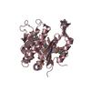
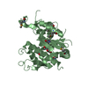
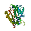
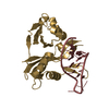
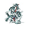
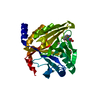
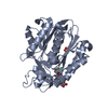
- PDB-3nro: Crystal Structure of putative transcriptional factor Lmo1026 from... -
+ Open data

- Basic information
| Entry | Database: PDB / ID: 3nro | ||||||
|---|---|---|---|---|---|---|---|
| Title | Crystal Structure of putative transcriptional factor Lmo1026 from Listeria monocytogenes (FRAGMENT 52-321), Northeast Structural Genomics Consortium Target LmR194 | ||||||
 Components Components | Lmo1026 protein | ||||||
 Keywords Keywords | structural genomics / unknown function / PSI-2 / Protein Structure Initiative / Northeast Structural Genomics Consortium / NESG | ||||||
| Function / homology | LCP protein / : / Cell envelope-related transcriptional attenuator domain / LytR_cpsA_psr family / Aminopeptidase / 3-Layer(aba) Sandwich / Alpha Beta / Lmo1026 protein Function and homology information Function and homology information | ||||||
| Biological species |  Listeria monocytogenes (bacteria) Listeria monocytogenes (bacteria) | ||||||
| Method |  X-RAY DIFFRACTION / SYNCHROTRON / SAD / Resolution: 2.9 Å X-RAY DIFFRACTION / SYNCHROTRON / SAD / Resolution: 2.9 Å | ||||||
 Authors Authors | Kuzin, A. / Su, M. / Seetharaman, J. / Mao, M. / Xiao, R. / Ciccosanti, C. / Lee, D. / Everett, J.K. / Nair, R. / Acton, T.B. ...Kuzin, A. / Su, M. / Seetharaman, J. / Mao, M. / Xiao, R. / Ciccosanti, C. / Lee, D. / Everett, J.K. / Nair, R. / Acton, T.B. / Rost, B. / Montelione, G.T. / Hunt, J.F. / Tong, L. / Northeast Structural Genomics Consortium (NESG) | ||||||
 Citation Citation | Journal: To be Published Title: Northeast Structural Genomics Consortium Target LmR194 Authors: Kuzin, A. / Su, M. / Seetharaman, J. / Mao, M. / Xiao, R. / Ciccosanti, C. / Lee, D. / Everett, J.K. / Nair, R. / Acton, T.B. / Rost, B. / Montelione, G.T. / Hunt, J.F. / Tong, L. | ||||||
| History |
|
- Structure visualization
| Structure viewer | Molecule: MolmilJmol/JSmol |
|---|
- Downloads & links
-Download
| PDBx/mmCIF format | 3nro.cif.gz | 191.9 KB | Display | PDBx/mmCIF format |
|---|---|---|---|---|
| PDB format | pdb3nro.ent.gz | 155.1 KB | Display | PDB format |
| PDBx/mmJSON format | 3nro.json.gz | Tree view | PDBx/mmJSON format | |
| Others |  Other downloads Other downloads |
-Validation report
| Arichive directory | https://data.pdbj.org/pub/pdb/validation_reports/nr/3nroftp://data.pdbj.org/pub/pdb/validation_reports/nr/3nro | HTTPS FTP |
|---|
-Related structure data
| Similar structure data | |
|---|---|
| Other databases |


-Links
 PDBj
PDBj- Assembly
| Deposited unit | 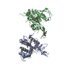
| ||||||||
|---|---|---|---|---|---|---|---|---|---|
| 1 | 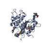
| ||||||||
| 2 | 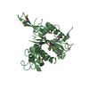
| ||||||||
| 3 | 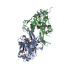
| ||||||||
| Unit cell |
| ||||||||
| Details | monomer,33.02 kD,91.5% |
-Components
| #1: Protein | Mass: 31287.561 Da / Num. of mol.: 2 Source method: isolated from a genetically manipulated source Source: (gene. exp.) Listeria monocytogenes (bacteria) / Gene: lmo1026 / Plasmid: pET 21-23C / Production host: Has protein modification | Y | |
|---|
-Experimental details
-Experiment
| Experiment | Method: X-RAY DIFFRACTION / Number of used crystals: 1 |
|---|
- Sample preparation
| Crystal | Density Matthews: 1.88 Å3/Da / Density % sol: 34.71 % |
|---|---|
| Crystal grow | Temperature: 293 K / pH: 8.5 Details: Protein solution: 100mM NaCl, 5mM DTT, 0.02% NaN3, 10mM Tris-HCl (pH 7.5) . Reservoir solution: LI2SO4 0.2M, Peg-4K 30%, 0.1M Tris, pH 8.5, macrobatch under oil, temperature 293K |
-Data collection
| Diffraction | Mean temperature: 100 K |
|---|---|
| Diffraction source | Source: SYNCHROTRON / Site: NSLS  / Beamline: X4A / Wavelength: 0.979 / Beamline: X4A / Wavelength: 0.979 |
| Detector | Type: ADSC QUANTUM 4 / Detector: CCD / Date: Jun 3, 2010 / Details: MIRRORS |
| Radiation | Monochromator: SI 111 CHANNEL / Protocol: SINGLE WAVELENGTH / Monochromatic (M) / Laue (L): M / Scattering type: x-ray |
| Radiation wavelength | Wavelength: 0.979 Å / Relative weight: 1 |
| Reflection | Resolution: 2.9→30 Å / Num. obs: 19967 / % possible obs: 98.9 % / Observed criterion σ(I): -3 / Redundancy: 3.8 % / Rsym value: 0.074 / Net I/σ(I): 19 |
- Processing
| Software |
| ||||||||||||||||||||||||||||||||||||||||
|---|---|---|---|---|---|---|---|---|---|---|---|---|---|---|---|---|---|---|---|---|---|---|---|---|---|---|---|---|---|---|---|---|---|---|---|---|---|---|---|---|---|
| Refinement | Method to determine structure: SAD / Resolution: 2.9→29.66 Å / Occupancy max: 1 / Occupancy min: 1 / SU ML: 0.39 / σ(F): 1.35 / Phase error: 25.6 / Stereochemistry target values: ML
| ||||||||||||||||||||||||||||||||||||||||
| Solvent computation | Shrinkage radii: 0.9 Å / VDW probe radii: 1.11 Å / Solvent model: FLAT BULK SOLVENT MODEL / Bsol: 45.59 Å2 / ksol: 0.32 e/Å3 | ||||||||||||||||||||||||||||||||||||||||
| Displacement parameters |
| ||||||||||||||||||||||||||||||||||||||||
| Refinement step | Cycle: LAST / Resolution: 2.9→29.66 Å
| ||||||||||||||||||||||||||||||||||||||||
| Refine LS restraints |
| ||||||||||||||||||||||||||||||||||||||||
| LS refinement shell |
| ||||||||||||||||||||||||||||||||||||||||
| Refinement TLS params. | Method: refined / Origin x: 20.3386 Å / Origin y: 35.6744 Å / Origin z: 45.1144 Å
| ||||||||||||||||||||||||||||||||||||||||
| Refinement TLS group | Selection details: ALL |
