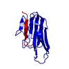
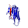
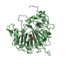
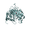
 Movie
Movie Controller


+ Open data

- Basic information
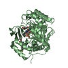
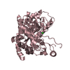
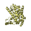
| Entry | Database: PDB / ID: 4en0 | |||||||||
|---|---|---|---|---|---|---|---|---|---|---|
| Title | Crystal structure of light | |||||||||
 Components Components | Tumor necrosis factor ligand superfamily member 14 | |||||||||
 Keywords Keywords | CYTOKINE / STRUCTURAL GENOMICS / PSI-BIOLOGY / NEW YORK STRUCTURAL GENOMICS RESEARCH CONSORTIUM / NYSGRC / IMMUNITY / TNF SUPERFAMILY / HVEM / DCR3 / N-GLYCOSYLATION / MEMBRANE / SECRETED PROTEIN / ATOMS-TO-ANIMALS: THE IMMUNE FUNCTION NETWORK / IFN / Jelly-roll Fold / Bind TNF receptor HVEM and LTbR / LTbR | |||||||||
| Function / homology |  Function and homology information Function and homology informationTNFs bind their physiological receptors / TNF receptor superfamily (TNFSF) members mediating non-canonical NF-kB pathway / positive regulation of T cell chemotaxis / T cell chemotaxis / cysteine-type endopeptidase inhibitor activity involved in apoptotic process / tumor necrosis factor receptor binding / positive regulation of extrinsic apoptotic signaling pathway / positive regulation of myoblast fusion / T cell homeostasis / T cell proliferation ...TNFs bind their physiological receptors / TNF receptor superfamily (TNFSF) members mediating non-canonical NF-kB pathway / positive regulation of T cell chemotaxis / T cell chemotaxis / cysteine-type endopeptidase inhibitor activity involved in apoptotic process / tumor necrosis factor receptor binding / positive regulation of extrinsic apoptotic signaling pathway / positive regulation of myoblast fusion / T cell homeostasis / T cell proliferation / positive regulation of myoblast differentiation / T cell costimulation / cytokine activity / T cell activation / TNFR2 non-canonical NF-kB pathway / cellular response to mechanical stimulus / positive regulation of non-canonical NF-kappaB signal transduction / positive regulation of canonical NF-kappaB signal transduction / cell surface receptor signaling pathway / immune response / signaling receptor binding / apoptotic process / signal transduction / : / identical protein binding / plasma membrane / cytoplasm Similarity search - Function | |||||||||
| Biological species |  Homo sapiens (human) Homo sapiens (human) | |||||||||
| Method |  X-RAY DIFFRACTION / SYNCHROTRON / MOLECULAR REPLACEMENT / Resolution: 2.59 Å X-RAY DIFFRACTION / SYNCHROTRON / MOLECULAR REPLACEMENT / Resolution: 2.59 Å | |||||||||
 Authors Authors | Zhan, C. / Liu, W. / Patskovsky, Y. / Ramagopal, U.A. / Bonanno, J.B. / Nathenson, S.G. / Almo, S.C. / New York Structural Genomics Research Consortium (NYSGRC) / Atoms-to-Animals: The Immune Function Network (IFN) | |||||||||
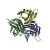
 Citation Citation | Journal: Structure / Year: 2014 Title: Mechanistic basis for functional promiscuity in the TNF and TNF receptor superfamilies: structure of the LIGHT:DcR3 assembly. Authors: Liu, W. / Zhan, C. / Cheng, H. / Kumar, P.R. / Bonanno, J.B. / Nathenson, S.G. / Almo, S.C. | |||||||||
| History |
|
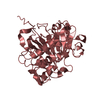
- Structure visualization
| Structure viewer | Molecule: MolmilJmol/JSmol |
|---|
- Downloads & links
-Download
| PDBx/mmCIF format | 4en0.cif.gz | 181.7 KB | Display | PDBx/mmCIF format |
|---|---|---|---|---|
| PDB format | pdb4en0.ent.gz | 145.8 KB | Display | PDB format |
| PDBx/mmJSON format | 4en0.json.gz | Tree view | PDBx/mmJSON format | |
| Others |  Other downloads Other downloads |
-Validation report
| Arichive directory | https://data.pdbj.org/pub/pdb/validation_reports/en/4en0ftp://data.pdbj.org/pub/pdb/validation_reports/en/4en0 | HTTPS FTP |
|---|
-Related structure data
| Related structure data | 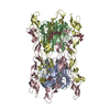 4j6gC 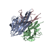 4kg8C 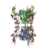 4kggC 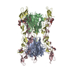 4kgqC  2qe3S S: Starting model for refinement C: citing same article ( |
|---|---|
| Similar structure data | |
| Other databases |
-Links
 PDBj
PDBj- Assembly
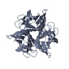
| Deposited unit | 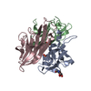
| ||||||||
|---|---|---|---|---|---|---|---|---|---|
| 1 |
| ||||||||
| Unit cell |
|
-Components
| #1: Protein | Mass: 18432.820 Da / Num. of mol.: 3 / Fragment: EXTRACELLULAR DOMAIN Source method: isolated from a genetically manipulated source Source: (gene. exp.) Homo sapiens (human) / Gene: HVEML, LIGHT, TNFSF14, UNQ391/PRO726 / Plasmid: PMT/BIP/V5-HIS / Production host:  #2: Chemical | ChemComp-PO4 / |   Mass: 94.971 Da / Num. of mol.: 1 / Source method: obtained synthetically / Formula: PO4 Mass: 94.971 Da / Num. of mol.: 1 / Source method: obtained synthetically / Formula: PO4#3: Chemical | ChemComp-GOL / |   Mass: 92.094 Da / Num. of mol.: 1 / Source method: obtained synthetically / Formula: C3H8O3 Mass: 92.094 Da / Num. of mol.: 1 / Source method: obtained synthetically / Formula: C3H8O3#4: Sugar | ChemComp-NAG / |   Type: D-saccharide, beta linking / Mass: 221.208 Da / Num. of mol.: 1 Type: D-saccharide, beta linking / Mass: 221.208 Da / Num. of mol.: 1Source method: isolated from a genetically manipulated source Formula: C8H15NO6 #5: Water | ChemComp-HOH / |  Mass: 18.015 Da / Num. of mol.: 12 / Source method: isolated from a natural source / Formula: H2O Mass: 18.015 Da / Num. of mol.: 12 / Source method: isolated from a natural source / Formula: H2OHas protein modification | Y | Sequence details | THE DIFFERENCE | |
|---|
-Experimental details
-Experiment
| Experiment | Method: X-RAY DIFFRACTION / Number of used crystals: 1 |
|---|
- Sample preparation
| Crystal | Density Matthews: 2.66 Å3/Da / Density % sol: 53.79 % |
|---|---|
| Crystal grow | Temperature: 290 K / Method: vapor diffusion, sitting drop / pH: 5.6 Details: 1.26M NAH2PO4, 0.14M K2HPO4 AND 0.2M NDSB-211, PH 5.6, VAPOR DIFFUSION, SITTING DROP, TEMPERATURE 290.0K |
-Data collection
| Diffraction | Mean temperature: 100 K |
|---|---|
| Diffraction source | Source: SYNCHROTRON / Site: NSLS  / Beamline: X29A / Wavelength: 0.9791 / Wavelength: 0.9791 Å / Beamline: X29A / Wavelength: 0.9791 / Wavelength: 0.9791 Å |
| Detector | Type: ADSC QUANTUM 315 / Detector: CCD / Date: Jan 28, 2011 |
| Radiation | Protocol: SINGLE WAVELENGTH / Monochromatic (M) / Laue (L): M / Scattering type: x-ray |
| Radiation wavelength | Wavelength: 0.9791 Å / Relative weight: 1 |
| Reflection | Resolution: 2.59→50 Å / Num. obs: 18664 / % possible obs: 100 % / Redundancy: 7.8 % / Biso Wilson estimate: 51.83 Å2 / Rmerge(I) obs: 0.136 / Rsym value: 0.09 / Net I/σ(I): 7.3 |
| Reflection shell | Resolution: 2.6→2.64 Å / Redundancy: 8.1 % / Rmerge(I) obs: 0.95 / Mean I/σ(I) obs: 2 / % possible all: 100 |
- Processing
| Software |
| ||||||||||||||||||||||||||||||||||||||||||||||||||||||||||||||||||||||||||||||||||||||||||||||||||||
|---|---|---|---|---|---|---|---|---|---|---|---|---|---|---|---|---|---|---|---|---|---|---|---|---|---|---|---|---|---|---|---|---|---|---|---|---|---|---|---|---|---|---|---|---|---|---|---|---|---|---|---|---|---|---|---|---|---|---|---|---|---|---|---|---|---|---|---|---|---|---|---|---|---|---|---|---|---|---|---|---|---|---|---|---|---|---|---|---|---|---|---|---|---|---|---|---|---|---|---|---|---|
| Refinement | Method to determine structure: MOLECULAR REPLACEMENT Starting model: PDB ENTRY 2QE3 Resolution: 2.59→46.122 Å / SU ML: 0.39 / σ(F): 0 / Phase error: 31.24 / Stereochemistry target values: ML
| ||||||||||||||||||||||||||||||||||||||||||||||||||||||||||||||||||||||||||||||||||||||||||||||||||||
| Solvent computation | Shrinkage radii: 0.73 Å / VDW probe radii: 1 Å / Solvent model: FLAT BULK SOLVENT MODEL / Bsol: 48.859 Å2 / ksol: 0.348 e/Å3 | ||||||||||||||||||||||||||||||||||||||||||||||||||||||||||||||||||||||||||||||||||||||||||||||||||||
| Displacement parameters |
| ||||||||||||||||||||||||||||||||||||||||||||||||||||||||||||||||||||||||||||||||||||||||||||||||||||
| Refinement step | Cycle: LAST / Resolution: 2.59→46.122 Å
| ||||||||||||||||||||||||||||||||||||||||||||||||||||||||||||||||||||||||||||||||||||||||||||||||||||
| Refine LS restraints |
| ||||||||||||||||||||||||||||||||||||||||||||||||||||||||||||||||||||||||||||||||||||||||||||||||||||
| LS refinement shell |
| ||||||||||||||||||||||||||||||||||||||||||||||||||||||||||||||||||||||||||||||||||||||||||||||||||||
| Refinement TLS params. | Method: refined / Refine-ID: X-RAY DIFFRACTION
| ||||||||||||||||||||||||||||||||||||||||||||||||||||||||||||||||||||||||||||||||||||||||||||||||||||
| Refinement TLS group |
|
