 Movie
Movie Controller

[English] 日本語
 Yorodumi
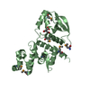
- PDB-4egq: Crystal structure of D-alanine-D-alanine ligase B from Burkholder... -
+ Open data

- Basic information
| Entry | Database: PDB / ID: 4egq | ||||||
|---|---|---|---|---|---|---|---|
| Title | Crystal structure of D-alanine-D-alanine ligase B from Burkholderia pseudomallei | ||||||
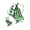
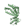
 Components Components | D-alanine--D-alanine ligase | ||||||
 Keywords Keywords | LIGASE / Structural Genomics / Seattle Structural Genomics Center for Infectious Disease / SSGCID / flexible protein / ATP-dependent / magnesium dependent / cell wall biogenesis | ||||||
| Function / homology |  Function and homology information Function and homology informationD-alanine-D-alanine ligase / D-alanine-D-alanine ligase activity / peptidoglycan biosynthetic process / cell wall organization / regulation of cell shape / ATP binding / metal ion binding / cytosol Similarity search - Function | ||||||
| Biological species |  Burkholderia pseudomallei (bacteria) Burkholderia pseudomallei (bacteria) | ||||||
| Method |  X-RAY DIFFRACTION / SYNCHROTRON / MOLECULAR REPLACEMENT / molecular replacement / Resolution: 2.2 Å X-RAY DIFFRACTION / SYNCHROTRON / MOLECULAR REPLACEMENT / molecular replacement / Resolution: 2.2 Å | ||||||
 Authors Authors | Seattle Structural Genomics Center for Infectious Disease (SSGCID) | ||||||
 Citation Citation | Journal: To be Published Title: Crystal structure of D-alanine-D-alanine ligase B from Burkholderia pseudomallei Authors: Edwards, T.E. / Fairman, J.W. / Abendroth, J. / Seattle Structural Genomics Center for Infectious Disease (SSGCID) | ||||||
| History |
|
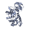
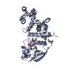
- Structure visualization
| Structure viewer | Molecule: MolmilJmol/JSmol |
|---|
- Downloads & links
-Download
| PDBx/mmCIF format | 4egq.cif.gz | 423.6 KB | Display | PDBx/mmCIF format |
|---|---|---|---|---|
| PDB format | pdb4egq.ent.gz | 346.3 KB | Display | PDB format |
| PDBx/mmJSON format | 4egq.json.gz | Tree view | PDBx/mmJSON format | |
| Others |  Other downloads Other downloads |
-Validation report
| Arichive directory | https://data.pdbj.org/pub/pdb/validation_reports/eg/4egqftp://data.pdbj.org/pub/pdb/validation_reports/eg/4egq | HTTPS FTP |
|---|
-Related structure data
| Related structure data | 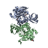 4eg0S S: Starting model for refinement |
|---|---|
| Similar structure data | |
| Other databases |


-Links
 PDBj
PDBj


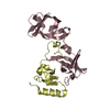
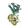
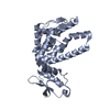
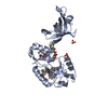
- Assembly
| Deposited unit | 
| ||||||||
|---|---|---|---|---|---|---|---|---|---|
| 1 | 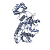
| ||||||||
| 2 | 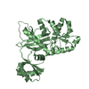
| ||||||||
| 3 | 
| ||||||||
| 4 | 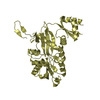
| ||||||||
| 5 | 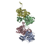
| ||||||||
| Unit cell |
|
-Components
| #1: Protein | Mass: 33677.227 Da / Num. of mol.: 4 Source method: isolated from a genetically manipulated source Source: (gene. exp.) Burkholderia pseudomallei (bacteria) / Strain: 1710b / Gene: BURPS1710b_3542, ddl / Production host: #2: Water | ChemComp-HOH / |  Mass: 18.015 Da / Num. of mol.: 193 / Source method: isolated from a natural source / Formula: H2O Mass: 18.015 Da / Num. of mol.: 193 / Source method: isolated from a natural source / Formula: H2O |
|---|
-Experimental details
-Experiment
| Experiment | Method: X-RAY DIFFRACTION / Number of used crystals: 1 |
|---|
- Sample preparation
| Crystal | Density Matthews: 2.14 Å3/Da / Density % sol: 42.64 % |
|---|---|
| Crystal grow | Temperature: 289 K / Method: vapor diffusion, sitting drop / pH: 9.5 Details: BupsA.00119.a.A1 PW25270 at 30.8 mg/mL against Wizard Full screen condition D2, 1 M NaK Tartrate, 0.1 M CHES pH 9.5, 0.2 M Li2SO4, crystal tracking ID 203378d2, VAPOR DIFFUSION, SITTING DROP, temperature 289K |
-Data collection
| Diffraction | Mean temperature: 100 K | |||||||||||||||||||||||||||||||||||||||||||||||||||||||||||||||||||||||||||||||||||||||||||||||||||||||||||||||||||||||||||||||||||||||||||||||||||
|---|---|---|---|---|---|---|---|---|---|---|---|---|---|---|---|---|---|---|---|---|---|---|---|---|---|---|---|---|---|---|---|---|---|---|---|---|---|---|---|---|---|---|---|---|---|---|---|---|---|---|---|---|---|---|---|---|---|---|---|---|---|---|---|---|---|---|---|---|---|---|---|---|---|---|---|---|---|---|---|---|---|---|---|---|---|---|---|---|---|---|---|---|---|---|---|---|---|---|---|---|---|---|---|---|---|---|---|---|---|---|---|---|---|---|---|---|---|---|---|---|---|---|---|---|---|---|---|---|---|---|---|---|---|---|---|---|---|---|---|---|---|---|---|---|---|---|---|---|
| Diffraction source | Source: SYNCHROTRON / Site: APS  / Beamline: 24-ID-E / Wavelength: 0.97946 Å / Beamline: 24-ID-E / Wavelength: 0.97946 Å | |||||||||||||||||||||||||||||||||||||||||||||||||||||||||||||||||||||||||||||||||||||||||||||||||||||||||||||||||||||||||||||||||||||||||||||||||||
| Detector | Date: Aug 1, 2008 | |||||||||||||||||||||||||||||||||||||||||||||||||||||||||||||||||||||||||||||||||||||||||||||||||||||||||||||||||||||||||||||||||||||||||||||||||||
| Radiation | Protocol: SINGLE WAVELENGTH / Monochromatic (M) / Laue (L): M / Scattering type: x-ray | |||||||||||||||||||||||||||||||||||||||||||||||||||||||||||||||||||||||||||||||||||||||||||||||||||||||||||||||||||||||||||||||||||||||||||||||||||
| Radiation wavelength | Wavelength: 0.97946 Å / Relative weight: 1 | |||||||||||||||||||||||||||||||||||||||||||||||||||||||||||||||||||||||||||||||||||||||||||||||||||||||||||||||||||||||||||||||||||||||||||||||||||
| Reflection | Resolution: 2.2→50 Å / Num. obs: 54908 / % possible obs: 97.8 % / Redundancy: 3.7 % / Rmerge(I) obs: 0.124 / Χ2: 1.001 / Net I/σ(I): 9.437 | |||||||||||||||||||||||||||||||||||||||||||||||||||||||||||||||||||||||||||||||||||||||||||||||||||||||||||||||||||||||||||||||||||||||||||||||||||
| Reflection shell |
|
-Phasing
| Phasing | Method: molecular replacement | |||||||||
|---|---|---|---|---|---|---|---|---|---|---|
| Phasing MR | Model details: Phaser MODE: MR_AUTO
|
- Processing
| Software |
| |||||||||||||||||||||||||||||||||||||||||||||||||||||||||||||||||||||||||||||||||||||||||||||||||||||||||||||||||||||||||||||
|---|---|---|---|---|---|---|---|---|---|---|---|---|---|---|---|---|---|---|---|---|---|---|---|---|---|---|---|---|---|---|---|---|---|---|---|---|---|---|---|---|---|---|---|---|---|---|---|---|---|---|---|---|---|---|---|---|---|---|---|---|---|---|---|---|---|---|---|---|---|---|---|---|---|---|---|---|---|---|---|---|---|---|---|---|---|---|---|---|---|---|---|---|---|---|---|---|---|---|---|---|---|---|---|---|---|---|---|---|---|---|---|---|---|---|---|---|---|---|---|---|---|---|---|---|---|---|
| Refinement | Method to determine structure: MOLECULAR REPLACEMENT Starting model: PDB entry 4EG0 Resolution: 2.2→35.27 Å / Cor.coef. Fo:Fc: 0.937 / Cor.coef. Fo:Fc free: 0.909 / WRfactor Rfree: 0.262 / WRfactor Rwork: 0.2191 / Occupancy max: 1 / Occupancy min: 1 / FOM work R set: 0.836 / SU B: 12.69 / SU ML: 0.157 / SU R Cruickshank DPI: 0.3313 / SU Rfree: 0.2304 / Cross valid method: THROUGHOUT / σ(F): 0 / ESU R: 0.331 / ESU R Free: 0.23 / Stereochemistry target values: MAXIMUM LIKELIHOOD Details: U VALUES: WITH TLS ADDED HYDROGENS HAVE BEEN ADDED IN THE RIDING POSITIONS NCS RESTRAINTS STATISTICS NCS TYPE: LOCAL NUMBER OF DIFFERENT NCS PAIRS: 6 GROUP CHAIN1 RANGE CHAIN2 RANGE COUNT ...Details: U VALUES: WITH TLS ADDED HYDROGENS HAVE BEEN ADDED IN THE RIDING POSITIONS NCS RESTRAINTS STATISTICS NCS TYPE: LOCAL NUMBER OF DIFFERENT NCS PAIRS: 6 GROUP CHAIN1 RANGE CHAIN2 RANGE COUNT RMS WEIGHT 1 A 5 311 B 5 311 8225 0.09 0.05 2 A 4 311 C 4 311 7892 0.13 0.05 3 A 4 311 D 4 311 7741 0.11 0.05 4 B 5 310 C 5 310 8154 0.13 0.05 5 B 5 311 D 5 311 8071 0.12 0.05 6 C 4 311 D 4 311 7824 0.12 0.05
| |||||||||||||||||||||||||||||||||||||||||||||||||||||||||||||||||||||||||||||||||||||||||||||||||||||||||||||||||||||||||||||
| Solvent computation | Ion probe radii: 0.8 Å / Shrinkage radii: 0.8 Å / VDW probe radii: 1.2 Å / Solvent model: MASK | |||||||||||||||||||||||||||||||||||||||||||||||||||||||||||||||||||||||||||||||||||||||||||||||||||||||||||||||||||||||||||||
| Displacement parameters | Biso max: 99.76 Å2 / Biso mean: 38.9073 Å2 / Biso min: 17.51 Å2
| |||||||||||||||||||||||||||||||||||||||||||||||||||||||||||||||||||||||||||||||||||||||||||||||||||||||||||||||||||||||||||||
| Refinement step | Cycle: LAST / Resolution: 2.2→35.27 Å
| |||||||||||||||||||||||||||||||||||||||||||||||||||||||||||||||||||||||||||||||||||||||||||||||||||||||||||||||||||||||||||||
| Refine LS restraints |
| |||||||||||||||||||||||||||||||||||||||||||||||||||||||||||||||||||||||||||||||||||||||||||||||||||||||||||||||||||||||||||||
| LS refinement shell | Resolution: 2.2→2.261 Å / Total num. of bins used: 20
| |||||||||||||||||||||||||||||||||||||||||||||||||||||||||||||||||||||||||||||||||||||||||||||||||||||||||||||||||||||||||||||
| Refinement TLS params. | Method: refined / Refine-ID: X-RAY DIFFRACTION
| |||||||||||||||||||||||||||||||||||||||||||||||||||||||||||||||||||||||||||||||||||||||||||||||||||||||||||||||||||||||||||||
| Refinement TLS group |
|
