 Movie
Movie Controller

[English] 日本語
 Yorodumi
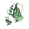
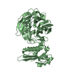
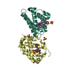
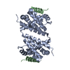
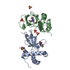
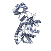
- PDB-3vba: Crystal structure of methanogen 3-isopropylmalate isomerase small... -
+ Open data

- Basic information
| Entry | Database: PDB / ID: 3vba | ||||||
|---|---|---|---|---|---|---|---|
| Title | Crystal structure of methanogen 3-isopropylmalate isomerase small subunit | ||||||
 Components Components | Isopropylmalate/citramalate isomerase small subunit | ||||||
 Keywords Keywords | LYASE / LeuD / cytosol | ||||||
| Function / homology |  Function and homology information Function and homology informationmaleate hydratase / (R)-2-methylmalate dehydratase / (R)-2-methylmalate dehydratase activity / maleate hydratase activity / 3-isopropylmalate dehydratase / 3-isopropylmalate dehydratase activity / L-leucine biosynthetic process / : Similarity search - Function | ||||||
| Biological species |   Methanocaldococcus jannaschii (archaea) Methanocaldococcus jannaschii (archaea) | ||||||
| Method |  X-RAY DIFFRACTION / SYNCHROTRON / MOLECULAR REPLACEMENT / Resolution: 2 Å X-RAY DIFFRACTION / SYNCHROTRON / MOLECULAR REPLACEMENT / Resolution: 2 Å | ||||||
 Authors Authors | Hwang, K.Y. / Lee, E.H. | ||||||
 Citation Citation | Journal: Biochem.Biophys.Res.Commun. / Year: 2012 Title: Crystal structure of LeuD from Methanococcus jannaschii. Authors: Lee, E.H. / Cho, Y.W. / Hwang, K.Y. | ||||||
| History |
|
- Structure visualization
| Structure viewer | Molecule: MolmilJmol/JSmol |
|---|
- Downloads & links
-Download
| PDBx/mmCIF format | 3vba.cif.gz | 209.3 KB | Display | PDBx/mmCIF format |
|---|---|---|---|---|
| PDB format | pdb3vba.ent.gz | 169.2 KB | Display | PDB format |
| PDBx/mmJSON format | 3vba.json.gz | Tree view | PDBx/mmJSON format | |
| Others |  Other downloads Other downloads |
-Validation report
| Arichive directory | https://data.pdbj.org/pub/pdb/validation_reports/vb/3vbaftp://data.pdbj.org/pub/pdb/validation_reports/vb/3vba | HTTPS FTP |
|---|
-Related structure data
| Similar structure data |
|---|


-Links
 PDBj
PDBj

- Assembly
| Deposited unit | 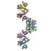
| ||||||||
|---|---|---|---|---|---|---|---|---|---|
| 1 | 
| ||||||||
| 2 | 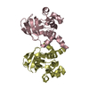
| ||||||||
| 3 | 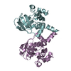
| ||||||||
| Unit cell |
|
-Components
| #1: Protein | Mass: 19471.545 Da / Num. of mol.: 6 Source method: isolated from a genetically manipulated source Source: (gene. exp.) Methanocaldococcus jannaschii (archaea)Strain: DSM 2661 / Gene: leuD, MJ1277 / Production host:  References: UniProt: Q58673, 3-isopropylmalate dehydratase, (R)-2-methylmalate dehydratase, maleate hydratase #2: Water | ChemComp-HOH / |  Mass: 18.015 Da / Num. of mol.: 721 / Source method: isolated from a natural source / Formula: H2O Mass: 18.015 Da / Num. of mol.: 721 / Source method: isolated from a natural source / Formula: H2O |
|---|
-Experimental details
-Experiment
| Experiment | Method: X-RAY DIFFRACTION / Number of used crystals: 1 |
|---|
- Sample preparation
| Crystal | Density Matthews: 2.24 Å3/Da / Density % sol: 45.08 % |
|---|---|
| Crystal grow | Temperature: 293 K / Method: vapor diffusion, hanging drop / pH: 7.9 Details: 25% PEG MME 2000, 0.1M Tris-HCl , pH 7.9, VAPOR DIFFUSION, HANGING DROP, temperature 293K |
-Data collection
| Diffraction | Mean temperature: 100 K |
|---|---|
| Diffraction source | Source: SYNCHROTRON / Site: SPring-8  / Beamline: BL26B2 / Wavelength: 1 Å / Beamline: BL26B2 / Wavelength: 1 Å |
| Detector | Type: msc saturna200 / Detector: CCD / Date: Apr 22, 2011 |
| Radiation | Monochromator: GRAPHITE / Protocol: SINGLE WAVELENGTH / Monochromatic (M) / Laue (L): M / Scattering type: x-ray |
| Radiation wavelength | Wavelength: 1 Å / Relative weight: 1 |
| Reflection | Resolution: 2→50 Å / Num. obs: 71994 / % possible obs: 98.6 % / Observed criterion σ(F): 2 / Observed criterion σ(I): 2 / Biso Wilson estimate: 19.45 Å2 |
| Reflection shell | Resolution: 1.98→2.03 Å / % possible all: 99.6 |
- Processing
| Software |
| |||||||||||||||||||||||||||||||||||||||||||||||||||||||||||||||||||||||||||||
|---|---|---|---|---|---|---|---|---|---|---|---|---|---|---|---|---|---|---|---|---|---|---|---|---|---|---|---|---|---|---|---|---|---|---|---|---|---|---|---|---|---|---|---|---|---|---|---|---|---|---|---|---|---|---|---|---|---|---|---|---|---|---|---|---|---|---|---|---|---|---|---|---|---|---|---|---|---|---|
| Refinement | Method to determine structure: MOLECULAR REPLACEMENT / Resolution: 2→43.895 Å / Occupancy max: 1 / Occupancy min: 1 / FOM work R set: 0.8467 / SU ML: 0.26 / σ(F): 0.09 / Phase error: 22.37 / Stereochemistry target values: ML
| |||||||||||||||||||||||||||||||||||||||||||||||||||||||||||||||||||||||||||||
| Solvent computation | Shrinkage radii: 0.9 Å / VDW probe radii: 1.11 Å / Solvent model: FLAT BULK SOLVENT MODEL / Bsol: 42.385 Å2 / ksol: 0.389 e/Å3 | |||||||||||||||||||||||||||||||||||||||||||||||||||||||||||||||||||||||||||||
| Displacement parameters | Biso max: 90.8 Å2 / Biso mean: 23.9456 Å2 / Biso min: 6.1 Å2
| |||||||||||||||||||||||||||||||||||||||||||||||||||||||||||||||||||||||||||||
| Refinement step | Cycle: LAST / Resolution: 2→43.895 Å
| |||||||||||||||||||||||||||||||||||||||||||||||||||||||||||||||||||||||||||||
| Refine LS restraints |
| |||||||||||||||||||||||||||||||||||||||||||||||||||||||||||||||||||||||||||||
| LS refinement shell | Refine-ID: X-RAY DIFFRACTION / Total num. of bins used: 10
|
