 Movie
Movie Controller


+ Open data

- Basic information
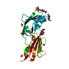
| Entry | Database: PDB / ID: 2zd2 | ||||||
|---|---|---|---|---|---|---|---|
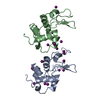
| Title | D202K mutant of P. denitrificans Atp12p | ||||||
 Components Components | ATP12 ATPase | ||||||
 Keywords Keywords | CHAPERONE / ATPase chaperone F1 assembly | ||||||
| Function / homology |  Function and homology information Function and homology information | ||||||
| Biological species |  Paracoccus denitrificans (bacteria) Paracoccus denitrificans (bacteria) | ||||||
| Method |  X-RAY DIFFRACTION / SYNCHROTRON / MOLECULAR REPLACEMENT / Resolution: 1.8 Å X-RAY DIFFRACTION / SYNCHROTRON / MOLECULAR REPLACEMENT / Resolution: 1.8 Å | ||||||
 Authors Authors | Gatti, D.L. / Ludlam, A. / Brunzelle, J. / Ackerman, S.H. | ||||||
 Citation Citation | Journal: J.Biol.Chem. / Year: 2009 Title: Chaperones of F1-ATPase Authors: Ludlam, A. / Brunzelle, J. / Pribyl, T. / Xu, X. / Gatti, D.L. / Ackerman, S.H. | ||||||
| History |
|
- Structure visualization
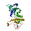
| Structure viewer | Molecule: MolmilJmol/JSmol |
|---|
- Downloads & links
-Download
| PDBx/mmCIF format | 2zd2.cif.gz | 115.6 KB | Display | PDBx/mmCIF format |
|---|---|---|---|---|
| PDB format | pdb2zd2.ent.gz | 87.9 KB | Display | PDB format |
| PDBx/mmJSON format | 2zd2.json.gz | Tree view | PDBx/mmJSON format | |
| Others |  Other downloads Other downloads |
-Validation report
| Arichive directory | https://data.pdbj.org/pub/pdb/validation_reports/zd/2zd2ftp://data.pdbj.org/pub/pdb/validation_reports/zd/2zd2 | HTTPS FTP |
|---|
-Related structure data
| Related structure data | 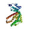 2r31C  2p4xS C: citing same article ( S: Starting model for refinement |
|---|---|
| Similar structure data |


-Links
 PDBj
PDBj
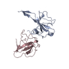
- Assembly
| Deposited unit | 
| ||||||||
|---|---|---|---|---|---|---|---|---|---|
| 1 | 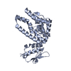
| ||||||||
| 2 | 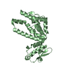
| ||||||||
| Unit cell |
|
-Components
| #1: Protein | Mass: 25798.365 Da / Num. of mol.: 2 / Mutation: D1202K, D2202K Source method: isolated from a genetically manipulated source Source: (gene. exp.) Paracoccus denitrificans (bacteria) / Strain: PD1222 / Gene: PDEN_0792 / Plasmid: PET28A-atp12D202K / Production host: #2: Water | ChemComp-HOH / |  Mass: 18.015 Da / Num. of mol.: 534 / Source method: isolated from a natural source / Formula: H2O Mass: 18.015 Da / Num. of mol.: 534 / Source method: isolated from a natural source / Formula: H2O |
|---|
-Experimental details
-Experiment
| Experiment | Method: X-RAY DIFFRACTION / Number of used crystals: 1 |
|---|
- Sample preparation
| Crystal | Density Matthews: 2.69 Å3/Da / Density % sol: 54.22 % |
|---|---|
| Crystal grow | Temperature: 278 K / Method: vapor diffusion, sitting drop / pH: 7.5 Details: 12% PEG 4000, 100mM Hepes-Na, pH 7.5, VAPOR DIFFUSION, SITTING DROP, temperature 278K |
-Data collection
| Diffraction | Mean temperature: 100 K |
|---|---|
| Diffraction source | Source: SYNCHROTRON / Site: APS  / Beamline: 21-ID-F / Wavelength: 0.97924 Å / Beamline: 21-ID-F / Wavelength: 0.97924 Å |
| Detector | Type: MARMOSAIC 300 mm CCD / Detector: CCD / Date: Oct 20, 2007 |
| Radiation | Monochromator: refractive beryllium lenses / Protocol: SINGLE WAVELENGTH / Monochromatic (M) / Laue (L): M / Scattering type: x-ray |
| Radiation wavelength | Wavelength: 0.97924 Å / Relative weight: 1 |
| Reflection | Resolution: 1.8→50 Å / Num. all: 48230 / Num. obs: 48230 / % possible obs: 97.5 % / Observed criterion σ(I): 0 / Redundancy: 3.8 % / Biso Wilson estimate: 20.7 Å2 / Rmerge(I) obs: 0.082 / Net I/σ(I): 15.3 |
| Reflection shell | Resolution: 1.8→1.83 Å / Redundancy: 3.1 % / Rmerge(I) obs: 0.555 / Mean I/σ(I) obs: 2.269 / Num. unique all: 2304 / % possible all: 95.6 |
- Processing
| Software |
| ||||||||||||||||||||||||||||||||||||||||||||||||||||||||||||||||||||||||||||||||
|---|---|---|---|---|---|---|---|---|---|---|---|---|---|---|---|---|---|---|---|---|---|---|---|---|---|---|---|---|---|---|---|---|---|---|---|---|---|---|---|---|---|---|---|---|---|---|---|---|---|---|---|---|---|---|---|---|---|---|---|---|---|---|---|---|---|---|---|---|---|---|---|---|---|---|---|---|---|---|---|---|---|
| Refinement | Method to determine structure: MOLECULAR REPLACEMENT Starting model: PDB ENTRY 2P4X Resolution: 1.8→35.21 Å / Rfactor Rfree error: 0.004 / Data cutoff high absF: 113038.34 / Data cutoff low absF: 0 / Isotropic thermal model: RESTRAINED / Cross valid method: THROUGHOUT / σ(F): 0 / Stereochemistry target values: Engh & Huber / Details: BULK SOLVENT MODEL USED
| ||||||||||||||||||||||||||||||||||||||||||||||||||||||||||||||||||||||||||||||||
| Solvent computation | Solvent model: FLAT MODEL / Bsol: 63.948 Å2 / ksol: 0.4 e/Å3 | ||||||||||||||||||||||||||||||||||||||||||||||||||||||||||||||||||||||||||||||||
| Displacement parameters | Biso mean: 28.8 Å2
| ||||||||||||||||||||||||||||||||||||||||||||||||||||||||||||||||||||||||||||||||
| Refine analyze |
| ||||||||||||||||||||||||||||||||||||||||||||||||||||||||||||||||||||||||||||||||
| Refinement step | Cycle: LAST / Resolution: 1.8→35.21 Å
| ||||||||||||||||||||||||||||||||||||||||||||||||||||||||||||||||||||||||||||||||
| Refine LS restraints |
| ||||||||||||||||||||||||||||||||||||||||||||||||||||||||||||||||||||||||||||||||
| LS refinement shell | Resolution: 1.8→1.91 Å / Rfactor Rfree error: 0.013 / Total num. of bins used: 6
| ||||||||||||||||||||||||||||||||||||||||||||||||||||||||||||||||||||||||||||||||
| Xplor file |
|
