 Movie
Movie Controller


+ Open data

- Basic information
| Entry | Database: PDB / ID: 4ee9 | ||||||
|---|---|---|---|---|---|---|---|
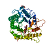
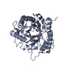
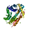
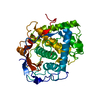
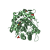
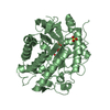
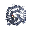
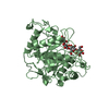
| Title | Crystal structure of the RBcel1 endo-1,4-glucanase | ||||||
 Components Components | Endoglucanase | ||||||
 Keywords Keywords | HYDROLASE / GLYCOSYL HYDROLASE FAMILY 5 / CELLULASE / TIM BARREL / BETA-1 / 4-ENDOGLUCANASE | ||||||
| Function / homology |  Function and homology information Function and homology information | ||||||
| Biological species |  uncultured bacterium (environmental samples) uncultured bacterium (environmental samples) | ||||||
| Method |  X-RAY DIFFRACTION / SYNCHROTRON / MOLECULAR REPLACEMENT / Resolution: 1.381 Å X-RAY DIFFRACTION / SYNCHROTRON / MOLECULAR REPLACEMENT / Resolution: 1.381 Å | ||||||
 Authors Authors | Delsaute, M. / Berlemont, R. / Van Elder, D. / Galleni, M. / Bauvois, C. | ||||||
 Citation Citation | Journal: Acta Crystallogr.,Sect.F / Year: 2013 Title: Three-dimensional structure of RBcel1, a metagenome-derived psychrotolerant family GH5 endoglucanase. Authors: Delsaute, M. / Berlemont, R. / Dehareng, D. / Van Elder, D. / Galleni, M. / Bauvois, C. #1: Journal: ISME J / Year: 2009 Title: Insights into bacterial cellulose biosynthesis by functional metagenomics on Antarctic soil samples. Authors: Berlemont, R. / Delsaute, M. / Pipers, D. / D'Amico, S. / Feller, G. / Galleni, M. / Power, P. | ||||||
| History |
|
- Structure visualization
| Structure viewer | Molecule: MolmilJmol/JSmol |
|---|
- Downloads & links
-Download
| PDBx/mmCIF format | 4ee9.cif.gz | 167.8 KB | Display | PDBx/mmCIF format |
|---|---|---|---|---|
| PDB format | pdb4ee9.ent.gz | 133.6 KB | Display | PDB format |
| PDBx/mmJSON format | 4ee9.json.gz | Tree view | PDBx/mmJSON format | |
| Others |  Other downloads Other downloads |
-Validation report
| Arichive directory | https://data.pdbj.org/pub/pdb/validation_reports/ee/4ee9ftp://data.pdbj.org/pub/pdb/validation_reports/ee/4ee9 | HTTPS FTP |
|---|
-Related structure data
| Similar structure data |
|---|


-Links
 PDBj
PDBj- Assembly
| Deposited unit | 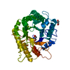
| ||||||||
|---|---|---|---|---|---|---|---|---|---|
| 1 |
| ||||||||
| Unit cell |
|
-Components
| #1: Protein | Mass: 36382.051 Da / Num. of mol.: 1 / Fragment: UNP residues 31-351 Source method: isolated from a genetically manipulated source Source: (gene. exp.) uncultured bacterium (environmental samples)Plasmid: pET-22b / Production host: | ||||
|---|---|---|---|---|---|
| #2: Chemical |   Mass: 122.143 Da / Num. of mol.: 3 / Source method: obtained synthetically / Formula: C4H12NO3 / Comment: pH buffer*YM Mass: 122.143 Da / Num. of mol.: 3 / Source method: obtained synthetically / Formula: C4H12NO3 / Comment: pH buffer*YM#3: Water | ChemComp-HOH / |  Mass: 18.015 Da / Num. of mol.: 557 / Source method: isolated from a natural source / Formula: H2O Mass: 18.015 Da / Num. of mol.: 557 / Source method: isolated from a natural source / Formula: H2OHas protein modification | Y | |
-Experimental details
-Experiment
| Experiment | Method: X-RAY DIFFRACTION / Number of used crystals: 1 |
|---|
- Sample preparation
| Crystal | Density Matthews: 2.24 Å3/Da / Density % sol: 45.05 % |
|---|---|
| Crystal grow | Temperature: 292 K / Method: vapor diffusion, hanging drop / pH: 6 Details: 17% PEG 600, 0.1M Tris-HCl pH 6.0, VAPOR DIFFUSION, HANGING DROP, temperature 292K |
-Data collection
| Diffraction | Mean temperature: 100 K |
|---|---|
| Diffraction source | Source: SYNCHROTRON / Site: ESRF  / Beamline: BM30A / Wavelength: 0.9797 Å / Beamline: BM30A / Wavelength: 0.9797 Å |
| Detector | Type: ADSC QUANTUM 315r / Detector: CCD / Date: Oct 2, 2011 |
| Radiation | Protocol: SINGLE WAVELENGTH / Monochromatic (M) / Laue (L): M / Scattering type: x-ray |
| Radiation wavelength | Wavelength: 0.9797 Å / Relative weight: 1 |
| Reflection | Resolution: 1.38→30.05 Å / Num. all: 67174 / Num. obs: 67782 / % possible obs: 99 % / Observed criterion σ(I): -3 / Redundancy: 9.5 % / Biso Wilson estimate: 11.9 Å2 / Rmerge(I) obs: 0.061 / Net I/σ(I): 20.5 |
| Reflection shell | Resolution: 1.38→1.46 Å / Redundancy: 8 % / Rmerge(I) obs: 0.64 / Mean I/σ(I) obs: 3.3 / Num. unique all: 9218 / % possible all: 94.5 |
- Processing
| Software |
| |||||||||||||||||||||||||||||||||||||||||||||||||||||||||||||||||||||||||||||||||||||||||||||||||||||||||||||||||||||||||||||||||||||||||||||||||||||||||||||||||||||||||||||||||||||||||||||||||||||||||||||||||||||||||
|---|---|---|---|---|---|---|---|---|---|---|---|---|---|---|---|---|---|---|---|---|---|---|---|---|---|---|---|---|---|---|---|---|---|---|---|---|---|---|---|---|---|---|---|---|---|---|---|---|---|---|---|---|---|---|---|---|---|---|---|---|---|---|---|---|---|---|---|---|---|---|---|---|---|---|---|---|---|---|---|---|---|---|---|---|---|---|---|---|---|---|---|---|---|---|---|---|---|---|---|---|---|---|---|---|---|---|---|---|---|---|---|---|---|---|---|---|---|---|---|---|---|---|---|---|---|---|---|---|---|---|---|---|---|---|---|---|---|---|---|---|---|---|---|---|---|---|---|---|---|---|---|---|---|---|---|---|---|---|---|---|---|---|---|---|---|---|---|---|---|---|---|---|---|---|---|---|---|---|---|---|---|---|---|---|---|---|---|---|---|---|---|---|---|---|---|---|---|---|---|---|---|---|---|---|---|---|---|---|---|---|---|---|---|---|---|---|---|---|
| Refinement | Method to determine structure: MOLECULAR REPLACEMENT / Resolution: 1.381→30.05 Å / SU ML: 0.13 / σ(F): 1.99 / Phase error: 18.12 / Stereochemistry target values: ML
| |||||||||||||||||||||||||||||||||||||||||||||||||||||||||||||||||||||||||||||||||||||||||||||||||||||||||||||||||||||||||||||||||||||||||||||||||||||||||||||||||||||||||||||||||||||||||||||||||||||||||||||||||||||||||
| Solvent computation | Shrinkage radii: 0.9 Å / VDW probe radii: 1.11 Å / Solvent model: FLAT BULK SOLVENT MODEL / Bsol: 62.101 Å2 / ksol: 0.376 e/Å3 | |||||||||||||||||||||||||||||||||||||||||||||||||||||||||||||||||||||||||||||||||||||||||||||||||||||||||||||||||||||||||||||||||||||||||||||||||||||||||||||||||||||||||||||||||||||||||||||||||||||||||||||||||||||||||
| Displacement parameters |
| |||||||||||||||||||||||||||||||||||||||||||||||||||||||||||||||||||||||||||||||||||||||||||||||||||||||||||||||||||||||||||||||||||||||||||||||||||||||||||||||||||||||||||||||||||||||||||||||||||||||||||||||||||||||||
| Refinement step | Cycle: LAST / Resolution: 1.381→30.05 Å
| |||||||||||||||||||||||||||||||||||||||||||||||||||||||||||||||||||||||||||||||||||||||||||||||||||||||||||||||||||||||||||||||||||||||||||||||||||||||||||||||||||||||||||||||||||||||||||||||||||||||||||||||||||||||||
| Refine LS restraints |
| |||||||||||||||||||||||||||||||||||||||||||||||||||||||||||||||||||||||||||||||||||||||||||||||||||||||||||||||||||||||||||||||||||||||||||||||||||||||||||||||||||||||||||||||||||||||||||||||||||||||||||||||||||||||||
| LS refinement shell |
|
