 Movie
Movie Controller

[English] 日本語
 Yorodumi
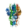
- PDB-4dw0: Crystal structure of the ATP-gated P2X4 ion channel in the closed... -
+ Open data

- Basic information
| Entry | Database: PDB / ID: 4dw0 | |||||||||
|---|---|---|---|---|---|---|---|---|---|---|
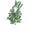
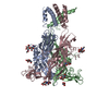
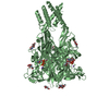
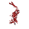
| Title | Crystal structure of the ATP-gated P2X4 ion channel in the closed, apo state at 2.9 Angstroms | |||||||||
 Components Components | P2X purinoceptor | |||||||||
 Keywords Keywords | TRANSPORT PROTEIN / ion channel | |||||||||
| Function / homology |  Function and homology information Function and homology informationElevation of cytosolic Ca2+ levels / Platelet homeostasis / purine nucleotide binding / extracellularly ATP-gated monoatomic cation channel activity / purinergic nucleotide receptor activity / transmembrane transporter complex / ATP-gated ion channel activity / ligand-gated monoatomic ion channel activity / CTP binding / response to ATP ...Elevation of cytosolic Ca2+ levels / Platelet homeostasis / purine nucleotide binding / extracellularly ATP-gated monoatomic cation channel activity / purinergic nucleotide receptor activity / transmembrane transporter complex / ATP-gated ion channel activity / ligand-gated monoatomic ion channel activity / CTP binding / response to ATP / monoatomic cation transport / monoatomic ion channel complex / calcium ion transport / postsynapse / lysosomal membrane / ATP binding / identical protein binding / membrane / plasma membrane Similarity search - Function | |||||||||
| Biological species |  | |||||||||
| Method |  X-RAY DIFFRACTION / SYNCHROTRON / MOLECULAR REPLACEMENT / Resolution: 2.9 Å X-RAY DIFFRACTION / SYNCHROTRON / MOLECULAR REPLACEMENT / Resolution: 2.9 Å | |||||||||
 Authors Authors | Hattori, M. / Gouaux, E. | |||||||||
 Citation Citation | Journal: Nature / Year: 2012 Title: Molecular mechanism of ATP binding and ion channel activation in P2X receptors. Authors: Hattori, M. / Gouaux, E. | |||||||||
| History |
|
- Structure visualization
| Structure viewer | Molecule: MolmilJmol/JSmol |
|---|
- Downloads & links
-Download
| PDBx/mmCIF format | 4dw0.cif.gz | 84.2 KB | Display | PDBx/mmCIF format |
|---|---|---|---|---|
| PDB format | pdb4dw0.ent.gz | 61.6 KB | Display | PDB format |
| PDBx/mmJSON format | 4dw0.json.gz | Tree view | PDBx/mmJSON format | |
| Others |  Other downloads Other downloads |
-Validation report
| Summary document | 4dw0_validation.pdf.gz | 750.7 KB | Display | wwPDB validaton report |
|---|---|---|---|---|
| Full document | 4dw0_full_validation.pdf.gz | 762.2 KB | Display | |
| Data in XML | 4dw0_validation.xml.gz | 10.9 KB | Display | |
| Data in CIF | 4dw0_validation.cif.gz | 15.5 KB | Display | |
| Arichive directory | https://data.pdbj.org/pub/pdb/validation_reports/dw/4dw0ftp://data.pdbj.org/pub/pdb/validation_reports/dw/4dw0 | HTTPS FTP |
-Related structure data


-Links
 PDBj
PDBj- Assembly
| Deposited unit | 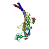
| ||||||||||||||||||
|---|---|---|---|---|---|---|---|---|---|---|---|---|---|---|---|---|---|---|---|
| 1 | 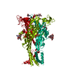
| ||||||||||||||||||
| Unit cell |
| ||||||||||||||||||
| Components on special symmetry positions |
| ||||||||||||||||||
| Details | The biological assembly is a trimer generated from the protomer in the asymmetric unit by the operations: -y, x-y, z and y-x, -x, z. |
-Components
-Protein , 1 types, 1 molecules A
| #1: Protein | Mass: 38161.656 Da / Num. of mol.: 1 / Fragment: UNP residues 28-381 / Mutation: C51F, N78K, N187R, H252R Source method: isolated from a genetically manipulated source Source: (gene. exp.)   Spodoptera frugiperda (fall armyworm) / Strain (production host): Sf9 / References: UniProt: Q6NYR1, UniProt: F8W463*PLUS Spodoptera frugiperda (fall armyworm) / Strain (production host): Sf9 / References: UniProt: Q6NYR1, UniProt: F8W463*PLUS |
|---|
-Sugars , 2 types, 2 molecules 
| #2: Polysaccharide | alpha-D-mannopyranose-(1-3)-beta-D-mannopyranose-(1-4)-2-acetamido-2-deoxy-beta-D-glucopyranose-(1- ...alpha-D-mannopyranose-(1-3)-beta-D-mannopyranose-(1-4)-2-acetamido-2-deoxy-beta-D-glucopyranose-(1-4)-2-acetamido-2-deoxy-beta-D-glucopyranose Source method: isolated from a genetically manipulated source |
|---|---|
| #3: Sugar | ChemComp-NAG /  Type: D-saccharide, beta linking / Mass: 221.208 Da / Num. of mol.: 1 Type: D-saccharide, beta linking / Mass: 221.208 Da / Num. of mol.: 1Source method: isolated from a genetically manipulated source Formula: C8H15NO6 |
-Non-polymers , 3 types, 84 molecules 


| #4: Chemical | ChemComp-GD /  Mass: 157.250 Da / Num. of mol.: 1 / Source method: obtained synthetically / Formula: Gd Mass: 157.250 Da / Num. of mol.: 1 / Source method: obtained synthetically / Formula: Gd | ||
|---|---|---|---|
| #5: Chemical | ChemComp-GOL /  Mass: 92.094 Da / Num. of mol.: 4 / Source method: obtained synthetically / Formula: C3H8O3 Mass: 92.094 Da / Num. of mol.: 4 / Source method: obtained synthetically / Formula: C3H8O3#6: Water | ChemComp-HOH / | Mass: 18.015 Da / Num. of mol.: 79 / Source method: isolated from a natural source / Formula: H2O |
-Details
| Has protein modification | Y |
|---|
-Experimental details
-Experiment
| Experiment | Method: X-RAY DIFFRACTION / Number of used crystals: 1 |
|---|
- Sample preparation
| Crystal | Density Matthews: 5.53 Å3/Da / Density % sol: 77.74 % |
|---|---|
| Crystal grow | Temperature: 277 K / Method: vapor diffusion, hanging drop / pH: 6.5 Details: 1 mM GdCl3, 20% PEG 3350, 100 mM MgCl2, 2M NaCl and 100 mM imidazole, pH 6.5, VAPOR DIFFUSION, HANGING DROP, temperature 277K |
-Data collection
| Diffraction | Mean temperature: 100 K |
|---|---|
| Diffraction source | Source: SYNCHROTRON / Site: APS  / Beamline: 24-ID-C / Wavelength: 0.97918 Å / Beamline: 24-ID-C / Wavelength: 0.97918 Å |
| Detector | Type: ADSC QUANTUM 315 / Detector: CCD / Date: Mar 10, 2011 |
| Radiation | Monochromator: Cryo-Cooled Si(111) double crystal / Protocol: SINGLE WAVELENGTH / Monochromatic (M) / Laue (L): M / Scattering type: x-ray |
| Radiation wavelength | Wavelength: 0.97918 Å / Relative weight: 1 |
| Reflection | Resolution: 2.9→50 Å / Num. all: 19275 / Num. obs: 18934 / % possible obs: 98.3 % / Observed criterion σ(I): -1 / Redundancy: 4.6 % |
| Reflection shell | Resolution: 2.9→2.95 Å / % possible all: 98.8 |
- Processing
| Software |
| ||||||||||||||||||||||||||||||||||||||||||||||||||||||||
|---|---|---|---|---|---|---|---|---|---|---|---|---|---|---|---|---|---|---|---|---|---|---|---|---|---|---|---|---|---|---|---|---|---|---|---|---|---|---|---|---|---|---|---|---|---|---|---|---|---|---|---|---|---|---|---|---|---|
| Refinement | Method to determine structure: MOLECULAR REPLACEMENT / Resolution: 2.9→47.184 Å / SU ML: 0.41 / σ(F): 0 / Phase error: 27.41 / Stereochemistry target values: ML Details: AUTHORS STATED THAT EVEN THOUGH THERE IS AN APPARENT GAP IN THE LATTICE, AUTHORS KNOW THAT THERE ARE STILL MANY AMINO ACID RESIDUES THAT AUTHORS HAVE NOT BEEN ABLE TO MODEL IN THE STRUCTURE ...Details: AUTHORS STATED THAT EVEN THOUGH THERE IS AN APPARENT GAP IN THE LATTICE, AUTHORS KNOW THAT THERE ARE STILL MANY AMINO ACID RESIDUES THAT AUTHORS HAVE NOT BEEN ABLE TO MODEL IN THE STRUCTURE AND WE BELIEVE THAT THESE RESIDUES ARE WHAT PROVIDE THE INTERACTIONS BETWEEN THE LAYERS
| ||||||||||||||||||||||||||||||||||||||||||||||||||||||||
| Solvent computation | Shrinkage radii: 0.72 Å / VDW probe radii: 1 Å / Solvent model: FLAT BULK SOLVENT MODEL / Bsol: 80 Å2 / ksol: 0.306 e/Å3 | ||||||||||||||||||||||||||||||||||||||||||||||||||||||||
| Displacement parameters |
| ||||||||||||||||||||||||||||||||||||||||||||||||||||||||
| Refinement step | Cycle: LAST / Resolution: 2.9→47.184 Å
| ||||||||||||||||||||||||||||||||||||||||||||||||||||||||
| Refine LS restraints |
| ||||||||||||||||||||||||||||||||||||||||||||||||||||||||
| LS refinement shell |
|
