 Movie
Movie Controller

[English] 日本語
 Yorodumi
- PDB-4dvc: Structural and functional studies of TcpG, the Vibrio cholerae Ds... -
+ Open data

- Basic information
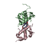
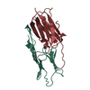
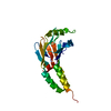
| Entry | Database: PDB / ID: 4dvc | ||||||
|---|---|---|---|---|---|---|---|
| Title | Structural and functional studies of TcpG, the Vibrio cholerae DsbA disulfide-forming protein required for pilus and cholera toxin production | ||||||
 Components Components | Thiol:disulfide interchange protein DsbA | ||||||
 Keywords Keywords | OXIDOREDUCTASE / Cholera / pilus assembly / Thioredoxin fold / DsbA-like / Disulfide bond / DsbB | ||||||
| Function / homology |  Function and homology information Function and homology informationprotein disulfide isomerase activity / cellular response to antibiotic / protein-disulfide reductase activity / outer membrane-bounded periplasmic space Similarity search - Function | ||||||
| Biological species |   Vibrio cholerae (bacteria) Vibrio cholerae (bacteria) | ||||||
| Method |  X-RAY DIFFRACTION / SYNCHROTRON / MOLECULAR REPLACEMENT / molecular replacement / Resolution: 1.2 Å X-RAY DIFFRACTION / SYNCHROTRON / MOLECULAR REPLACEMENT / molecular replacement / Resolution: 1.2 Å | ||||||
 Authors Authors | Walden, P.M. / Martin, J.L. | ||||||
 Citation Citation | Journal: Acta Crystallogr.,Sect.D / Year: 2012 Title: The 1.2 A resolution crystal structure of TcpG, the Vibrio cholerae DsbA disulfide-forming protein required for pilus and cholera-toxin production Authors: Walden, P.M. / Heras, B. / Chen, K.-E. / Halili, M.A. / Rimmer, K. / Sharma, P. / Scanlon, M.J. / Martin, J.L. | ||||||
| History |
|
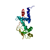
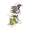
- Structure visualization
| Structure viewer | Molecule: MolmilJmol/JSmol |
|---|
- Downloads & links
-Download
| PDBx/mmCIF format | 4dvc.cif.gz | 131.8 KB | Display | PDBx/mmCIF format |
|---|---|---|---|---|
| PDB format | pdb4dvc.ent.gz | 105.4 KB | Display | PDB format |
| PDBx/mmJSON format | 4dvc.json.gz | Tree view | PDBx/mmJSON format | |
| Others |  Other downloads Other downloads |
-Validation report
| Arichive directory | https://data.pdbj.org/pub/pdb/validation_reports/dv/4dvcftp://data.pdbj.org/pub/pdb/validation_reports/dv/4dvc | HTTPS FTP |
|---|
-Related structure data
| Similar structure data |
|---|


-Links
 PDBj
PDBj- Assembly
| Deposited unit | 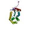
| ||||||||||||||||||
|---|---|---|---|---|---|---|---|---|---|---|---|---|---|---|---|---|---|---|---|
| 1 |
| ||||||||||||||||||
| Unit cell |
| ||||||||||||||||||
| Components on special symmetry positions |
|
-Components
| #1: Protein | Mass: 20748.580 Da / Num. of mol.: 1 / Fragment: UNP residues 19-200 / Mutation: R134H, V164A Source method: isolated from a genetically manipulated source Source: (gene. exp.) Vibrio cholerae (bacteria) / Strain: serotype O1 (ATCC 39315 / El Tor Inaba N16961) / Gene: tpcG / Plasmid: pET-21a / Production host: | ||||||||
|---|---|---|---|---|---|---|---|---|---|
| #2: Chemical |   Mass: 78.133 Da / Num. of mol.: 2 / Source method: obtained synthetically / Formula: C2H6OS / Comment: DMSO, precipitant*YM Mass: 78.133 Da / Num. of mol.: 2 / Source method: obtained synthetically / Formula: C2H6OS / Comment: DMSO, precipitant*YM#3: Chemical | ChemComp-SO4 / |   Mass: 96.063 Da / Num. of mol.: 1 / Source method: obtained synthetically / Formula: SO4 Mass: 96.063 Da / Num. of mol.: 1 / Source method: obtained synthetically / Formula: SO4#4: Water | ChemComp-HOH / |  Mass: 18.015 Da / Num. of mol.: 284 / Source method: isolated from a natural source / Formula: H2O Mass: 18.015 Da / Num. of mol.: 284 / Source method: isolated from a natural source / Formula: H2OHas protein modification | Y | Sequence details | THIS SEQUENCE CONFLICT IS BASED ON REFERENCE 1 OF DATABASE UNIPROTKB/SWISS-PROT P32557 (DSBA_VIBCH). | |
-Experimental details
-Experiment
| Experiment | Method: X-RAY DIFFRACTION / Number of used crystals: 1 |
|---|
- Sample preparation
| Crystal | Density Matthews: 2.17 Å3/Da / Density % sol: 43.36 % |
|---|---|
| Crystal grow | Temperature: 277.15 K / Method: hanging drop / pH: 6.4 Details: PEG 4000, MES, pH 6.4, hanging drop, temperature 277.15K |
-Data collection
| Diffraction | Mean temperature: 100 K |
|---|---|
| Diffraction source | Source: SYNCHROTRON / Site: Australian Synchrotron  / Beamline: MX2 / Wavelength: 0.9536 Å / Beamline: MX2 / Wavelength: 0.9536 Å |
| Detector | Type: ADSC QUANTUM 315r / Detector: CCD / Date: Jul 9, 2009 |
| Radiation | Monochromator: Sagitally focussed Si / Protocol: SINGLE WAVELENGTH / Monochromatic (M) / Laue (L): M / Scattering type: x-ray |
| Radiation wavelength | Wavelength: 0.9536 Å / Relative weight: 1 |
| Reflection | Resolution: 1.2→64.2 Å / Num. obs: 56644 / % possible obs: 99.7 % / Observed criterion σ(F): 0 / Observed criterion σ(I): 0 / Redundancy: 14 % / Biso Wilson estimate: 10 Å2 / Rmerge(I) obs: 0.103 / Net I/σ(I): 18 |
| Reflection shell | Highest resolution: 1.2 Å / Redundancy: 12.9 % / Rmerge(I) obs: 0.103 / Mean I/σ(I) obs: 3.1 / % possible all: 97.4 |
-Phasing
| Phasing | Method: molecular replacement |
|---|
- Processing
| Software |
| ||||||||||||||||||||||||||||||||||||||||||||||||||||||||||||||||||||||||||||||||||||||||||
|---|---|---|---|---|---|---|---|---|---|---|---|---|---|---|---|---|---|---|---|---|---|---|---|---|---|---|---|---|---|---|---|---|---|---|---|---|---|---|---|---|---|---|---|---|---|---|---|---|---|---|---|---|---|---|---|---|---|---|---|---|---|---|---|---|---|---|---|---|---|---|---|---|---|---|---|---|---|---|---|---|---|---|---|---|---|---|---|---|---|---|---|
| Refinement | Method to determine structure: MOLECULAR REPLACEMENT / Resolution: 1.2→32.132 Å / Occupancy max: 1 / Occupancy min: 0 / SU ML: 0.09 / σ(F): 1.34 / Phase error: 11.33 / Stereochemistry target values: MLHL
| ||||||||||||||||||||||||||||||||||||||||||||||||||||||||||||||||||||||||||||||||||||||||||
| Solvent computation | Shrinkage radii: 0.9 Å / VDW probe radii: 1.11 Å / Solvent model: FLAT BULK SOLVENT MODEL | ||||||||||||||||||||||||||||||||||||||||||||||||||||||||||||||||||||||||||||||||||||||||||
| Displacement parameters | Biso mean: 16.3622 Å2
| ||||||||||||||||||||||||||||||||||||||||||||||||||||||||||||||||||||||||||||||||||||||||||
| Refinement step | Cycle: LAST / Resolution: 1.2→32.132 Å
| ||||||||||||||||||||||||||||||||||||||||||||||||||||||||||||||||||||||||||||||||||||||||||
| Refine LS restraints |
| ||||||||||||||||||||||||||||||||||||||||||||||||||||||||||||||||||||||||||||||||||||||||||
| LS refinement shell | Refine-ID: X-RAY DIFFRACTION / Total num. of bins used: 14
|
