 Movie
Movie Controller

[English] 日本語
 Yorodumi
- PDB-4de7: Crystal structure of glucosyl-3-phosphoglycerate synthase from My... -
+ Open data

- Basic information
| Entry | Database: PDB / ID: 4de7 | ||||||
|---|---|---|---|---|---|---|---|
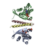
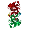
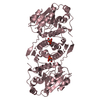
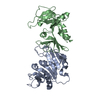
| Title | Crystal structure of glucosyl-3-phosphoglycerate synthase from Mycobacterium tuberculosis in complex with Mg2+ and uridine-diphosphate (UDP) | ||||||
 Components Components | GLUCOSYL-3-PHOSPHOGLYCERATE SYNTHASE (GpgS) | ||||||
 Keywords Keywords | TRANSFERASE | ||||||
| Function / homology |  Function and homology information Function and homology informationglucosyl-3-phosphoglycerate synthase / UDP-alpha-D-glucose metabolic process / hexosyltransferase activity / glycosyltransferase activity / magnesium ion binding / protein homodimerization activity / metal ion binding Similarity search - Function | ||||||
| Biological species |   Mycobacterium tuberculosis (bacteria) Mycobacterium tuberculosis (bacteria) | ||||||
| Method |  X-RAY DIFFRACTION / SYNCHROTRON / MOLECULAR REPLACEMENT / Resolution: 3 Å X-RAY DIFFRACTION / SYNCHROTRON / MOLECULAR REPLACEMENT / Resolution: 3 Å | ||||||
 Authors Authors | Albesa-Jove, D. / Urresti, S. / van der Woerd, M. / Guerin, M.E. | ||||||
 Citation Citation | Journal: J.Biol.Chem. / Year: 2012 Title: Mechanistic insights into the retaining glucosyl-3-phosphoglycerate synthase from mycobacteria. Authors: Urresti, S. / Albesa-Jove, D. / Schaeffer, F. / Pham, H.T. / Kaur, D. / Gest, P. / van der Woerd, M.J. / Carreras-Gonzalez, A. / Lopez-Fernandez, S. / Alzari, P.M. / Brennan, P.J. / Jackson, M. / Guerin, M.E. | ||||||
| History |
|
- Structure visualization
| Structure viewer | Molecule: MolmilJmol/JSmol |
|---|
- Downloads & links
-Download
| PDBx/mmCIF format | 4de7.cif.gz | 70 KB | Display | PDBx/mmCIF format |
|---|---|---|---|---|
| PDB format | pdb4de7.ent.gz | 50.2 KB | Display | PDB format |
| PDBx/mmJSON format | 4de7.json.gz | Tree view | PDBx/mmJSON format | |
| Others |  Other downloads Other downloads |
-Validation report
| Arichive directory | https://data.pdbj.org/pub/pdb/validation_reports/de/4de7ftp://data.pdbj.org/pub/pdb/validation_reports/de/4de7 | HTTPS FTP |
|---|
-Related structure data
-Links
 PDBj
PDBj


- Assembly
| Deposited unit | 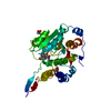
| ||||||||
|---|---|---|---|---|---|---|---|---|---|
| 1 | 
| ||||||||
| Unit cell |
|
-Components
| #1: Protein | Mass: 36582.656 Da / Num. of mol.: 1 Source method: isolated from a genetically manipulated source Source: (gene. exp.) Mycobacterium tuberculosis (bacteria) / Strain: H37Rv / Gene: gpgS, MT1246, Rv1208 / Plasmid: pET28a-gpgS / Production host: References: UniProt: O05309, UniProt: P9WMW9*PLUS, Transferases; Glycosyltransferases; Hexosyltransferases | ||
|---|---|---|---|
| #2: Chemical | ChemComp-UDP /   Type: RNA linking / Mass: 404.161 Da / Num. of mol.: 1 / Source method: obtained synthetically / Formula: C9H14N2O12P2 / Comment: UDP*YM Type: RNA linking / Mass: 404.161 Da / Num. of mol.: 1 / Source method: obtained synthetically / Formula: C9H14N2O12P2 / Comment: UDP*YM | ||
| #3: Chemical |   Mass: 92.094 Da / Num. of mol.: 2 / Source method: obtained synthetically / Formula: C3H8O3 Mass: 92.094 Da / Num. of mol.: 2 / Source method: obtained synthetically / Formula: C3H8O3#4: Water | ChemComp-HOH / |  Mass: 18.015 Da / Num. of mol.: 22 / Source method: isolated from a natural source / Formula: H2O Mass: 18.015 Da / Num. of mol.: 22 / Source method: isolated from a natural source / Formula: H2O |
-Experimental details
-Experiment
| Experiment | Method: X-RAY DIFFRACTION / Number of used crystals: 1 |
|---|
- Sample preparation
| Crystal | Density Matthews: 4.26 Å3/Da / Density % sol: 71.14 % |
|---|---|
| Crystal grow | Temperature: 289 K / Method: vapor diffusion, sitting drop / pH: 8 Details: 1 mM MgCl2, 5mM UDP, 1.5M NaCl, 10%(v/v) ethanol, Tris-HCL pH 8.0, VAPOR DIFFUSION, SITTING DROP, temperature 289K |
-Data collection
| Diffraction | Mean temperature: 100 K |
|---|---|
| Diffraction source | Source: SYNCHROTRON / Site: ALS  / Beamline: 4.2.2 / Wavelength: 1 Å / Beamline: 4.2.2 / Wavelength: 1 Å |
| Detector | Type: NOIR-1 / Detector: CCD / Date: Jul 22, 2008 |
| Radiation | Monochromator: Rosenbaum-Rock Si(111) sagitally focused monochromator Protocol: SINGLE WAVELENGTH / Monochromatic (M) / Laue (L): M / Scattering type: x-ray |
| Radiation wavelength | Wavelength: 1 Å / Relative weight: 1 |
| Reflection | Resolution: 3→41.58 Å / Num. all: 12217 / Num. obs: 12217 / % possible obs: 100 % / Observed criterion σ(F): 0 / Observed criterion σ(I): 0 / Redundancy: 7.22 % / Biso Wilson estimate: 68.5 Å2 / Rmerge(I) obs: 0.107 / Net I/σ(I): 10.3 |
| Reflection shell | Resolution: 3→3.11 Å / Redundancy: 7.08 % / Rmerge(I) obs: 0.413 / Mean I/σ(I) obs: 3.3 / Num. unique all: 1205 / % possible all: 100 |
- Processing
| Software |
| |||||||||||||||||||||||||||||||||||
|---|---|---|---|---|---|---|---|---|---|---|---|---|---|---|---|---|---|---|---|---|---|---|---|---|---|---|---|---|---|---|---|---|---|---|---|---|
| Refinement | Method to determine structure: MOLECULAR REPLACEMENT / Resolution: 3→39.077 Å / SU ML: 0.85 / σ(F): 1.46 / Phase error: 23.89 / Stereochemistry target values: ML Details: AUTHORS STATE THE FOLLOWING ON THE DENSITY ON UDP. THE DENSITY IS VERY LOW, BUT WE DO BELIEVE THE LIGAND IS PRESENT. WE THINK THAT THE LOW OCCUPANCY AND THE PARTIAL DISORDER OF UDP MOLECULE ...Details: AUTHORS STATE THE FOLLOWING ON THE DENSITY ON UDP. THE DENSITY IS VERY LOW, BUT WE DO BELIEVE THE LIGAND IS PRESENT. WE THINK THAT THE LOW OCCUPANCY AND THE PARTIAL DISORDER OF UDP MOLECULE RESULTS IN VERY WEEK ELECTRON DENSITY. BOTH, THE ALPHA- AND BETA-PHOSPHATES OF UDP ARE DISORDERED, AND NO ELECTRON DENSITY IS VISIBLE FOR THEM. WE BELIEVE THE DISORDER OF PHOSPHATE GROUPS IS DUE TO THE ABSENCE OF DIVALENT CATION BOND TO THE STRUCTURE, WHICH USUALLY COORDINATES BETWEEN THE PHOSPHATE GROUPS AND THE ENZYME. COORDINATION OF THE DIVALENT CATION IS PH DEPENDENT, AT PH < 6 THE RESIDUE HIS258 IS FOUND PROTONATED, AND IS UNABLE TO COORDINATE DIVALENT CATIONS. WE BELIEVE THAT IS WHAT HAPPENS IN THIS CASE, RESULTING IN PARTIAL DISORDER OF UDP AND LOW OCCUPANCY. NONETHELESS, WEEK ELECTRON DENSITY IS FOUND FOR THE URIDINE MOIETY, SO WE FELL IT IS STILL INFORMATIVE TO INCLUDE IT IN THE STRUCTURE.
| |||||||||||||||||||||||||||||||||||
| Solvent computation | Shrinkage radii: 0.86 Å / VDW probe radii: 1.1 Å / Solvent model: FLAT BULK SOLVENT MODEL / Bsol: 38.265 Å2 / ksol: 0.345 e/Å3 | |||||||||||||||||||||||||||||||||||
| Displacement parameters |
| |||||||||||||||||||||||||||||||||||
| Refinement step | Cycle: LAST / Resolution: 3→39.077 Å
| |||||||||||||||||||||||||||||||||||
| Refine LS restraints |
| |||||||||||||||||||||||||||||||||||
| LS refinement shell |
|
