 Movie
Movie Controller

[English] 日本語
 Yorodumi
- PDB-4ha9: Structural insights into the reduction mechanism of Saccharomyces... -
+ Open data

- Basic information
| Entry | Database: PDB / ID: 4ha9 | ||||||
|---|---|---|---|---|---|---|---|
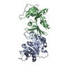
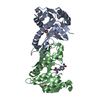
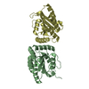
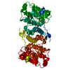
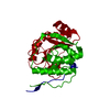
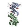
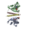
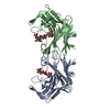
| Title | Structural insights into the reduction mechanism of Saccharomyces cerevisia Riboflavin Biosynthesis Reductase Rib7 | ||||||
 Components Components | 2,5-diamino-6-ribosylamino-4(3H)-pyrimidinone 5'-phosphate reductase | ||||||
 Keywords Keywords | OXIDOREDUCTASE / reductase / NADPH binding | ||||||
| Function / homology |  Function and homology information Function and homology information2,5-diamino-6-(ribosylamino)-4(3H)-pyrimidinone 5'-phosphate reductase / 5-amino-6-(5-phosphoribosylamino)uracil reductase activity / riboflavin biosynthetic process / NADP binding Similarity search - Function | ||||||
| Biological species |  | ||||||
| Method |  X-RAY DIFFRACTION / SYNCHROTRON / molecular replacement / Resolution: 2.35 Å X-RAY DIFFRACTION / SYNCHROTRON / molecular replacement / Resolution: 2.35 Å | ||||||
 Authors Authors | Lv, Z. / Sun, J. / Liu, Y. | ||||||
 Citation Citation | Journal: Plos One / Year: 2013 Title: Structural and functional insights into Saccharomyces cerevisiae riboflavin biosynthesis reductase RIB7. Authors: Lv, Z. / Sun, J. / Liu, Y. | ||||||
| History |
|
- Structure visualization
| Structure viewer | Molecule: MolmilJmol/JSmol |
|---|
- Downloads & links
-Download
| PDBx/mmCIF format | 4ha9.cif.gz | 185.3 KB | Display | PDBx/mmCIF format |
|---|---|---|---|---|
| PDB format | pdb4ha9.ent.gz | 148 KB | Display | PDB format |
| PDBx/mmJSON format | 4ha9.json.gz | Tree view | PDBx/mmJSON format | |
| Others |  Other downloads Other downloads |
-Validation report
| Arichive directory | https://data.pdbj.org/pub/pdb/validation_reports/ha/4ha9ftp://data.pdbj.org/pub/pdb/validation_reports/ha/4ha9 | HTTPS FTP |
|---|
-Related structure data


-Links
 PDBj
PDBj

- Assembly
| Deposited unit | 
| ||||||||
|---|---|---|---|---|---|---|---|---|---|
| 1 |
| ||||||||
| Unit cell |
|
-Components
| #1: Protein | Mass: 27557.598 Da / Num. of mol.: 2 Source method: isolated from a genetically manipulated source Source: (gene. exp.) Strain: ATCC 204508 / S288c / Gene: RIB7, YBR153W, YBR1203 / Production host:  References: UniProt: P33312, 2,5-diamino-6-(ribosylamino)-4(3H)-pyrimidinone 5'-phosphate reductase #2: Chemical | ChemComp-NDP / |   Mass: 745.421 Da / Num. of mol.: 1 / Source method: obtained synthetically / Formula: C21H30N7O17P3 Mass: 745.421 Da / Num. of mol.: 1 / Source method: obtained synthetically / Formula: C21H30N7O17P3#3: Water | ChemComp-HOH / |  Mass: 18.015 Da / Num. of mol.: 70 / Source method: isolated from a natural source / Formula: H2O Mass: 18.015 Da / Num. of mol.: 70 / Source method: isolated from a natural source / Formula: H2O |
|---|
-Experimental details
-Experiment
| Experiment | Method: X-RAY DIFFRACTION / Number of used crystals: 1 |
|---|
- Sample preparation
| Crystal | Density Matthews: 2.16 Å3/Da / Density % sol: 43.03 % |
|---|---|
| Crystal grow | Temperature: 289 K / Method: vapor diffusion, hanging drop / pH: 6.5 Details: 0.05M Calcium chloride dihydrate, 0.1M BIS-TRIS, 30% v/v Polyethylene glycol monomethyl ether 550 , pH 6.5, VAPOR DIFFUSION, HANGING DROP, temperature 289K |
-Data collection
| Diffraction | Mean temperature: 100 K |
|---|---|
| Diffraction source | Source: SYNCHROTRON / Site: Australian Synchrotron  / Beamline: MX1 / Wavelength: 0.9184 Å / Beamline: MX1 / Wavelength: 0.9184 Å |
| Detector | Type: ADSC QUANTUM 210 / Detector: CCD / Date: Jun 21, 2012 |
| Radiation | Protocol: SINGLE WAVELENGTH / Monochromatic (M) / Laue (L): M / Scattering type: x-ray |
| Radiation wavelength | Wavelength: 0.9184 Å / Relative weight: 1 |
| Reflection | Resolution: 2.35→34.43 Å / Num. all: 20528 / Num. obs: 20487 / % possible obs: 99.8 % / Observed criterion σ(F): 1.4 / Observed criterion σ(I): 2 |
| Reflection shell | Resolution: 2.35→2.39 Å / % possible all: 100 |
- Processing
| Software |
| ||||||||||||||||||||||||||||||||||||||||||||||||||||||||
|---|---|---|---|---|---|---|---|---|---|---|---|---|---|---|---|---|---|---|---|---|---|---|---|---|---|---|---|---|---|---|---|---|---|---|---|---|---|---|---|---|---|---|---|---|---|---|---|---|---|---|---|---|---|---|---|---|---|
| Refinement | Method to determine structure: molecular replacement / Resolution: 2.35→34.43 Å / SU ML: 0.36 / σ(F): 1.34 / Phase error: 28.07 / Stereochemistry target values: ML
| ||||||||||||||||||||||||||||||||||||||||||||||||||||||||
| Solvent computation | Shrinkage radii: 0.98 Å / VDW probe radii: 1.2 Å / Solvent model: FLAT BULK SOLVENT MODEL / Bsol: 43.483 Å2 / ksol: 0.342 e/Å3 | ||||||||||||||||||||||||||||||||||||||||||||||||||||||||
| Displacement parameters |
| ||||||||||||||||||||||||||||||||||||||||||||||||||||||||
| Refinement step | Cycle: LAST / Resolution: 2.35→34.43 Å
| ||||||||||||||||||||||||||||||||||||||||||||||||||||||||
| Refine LS restraints |
| ||||||||||||||||||||||||||||||||||||||||||||||||||||||||
| LS refinement shell |
| ||||||||||||||||||||||||||||||||||||||||||||||||||||||||
| Refinement TLS params. | Method: refined / Origin x: -10.8081 Å / Origin y: 10.3548 Å / Origin z: -18.1256 Å
| ||||||||||||||||||||||||||||||||||||||||||||||||||||||||
| Refinement TLS group | Selection details: all |
