 Movie
Movie Controller

[English] 日本語
 Yorodumi
- PDB-3e25: Crystal structure of M. tuberculosis glucosyl-3-phosphoglycerate ... -
+ Open data

- Basic information
| Entry | Database: PDB / ID: 3.0E+25 | ||||||
|---|---|---|---|---|---|---|---|
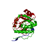
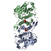
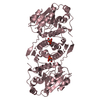
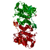
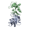
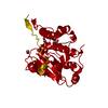
| Title | Crystal structure of M. tuberculosis glucosyl-3-phosphoglycerate synthase | ||||||
 Components Components | Putative uncharacterized protein | ||||||
 Keywords Keywords | TRANSFERASE / glucosyltransferase / mycobacterial / GT81 UDP-glucose / 3-phosphoglycerate | ||||||
| Function / homology |  Function and homology information Function and homology informationglucosyl-3-phosphoglycerate synthase / UDP-alpha-D-glucose metabolic process / hexosyltransferase activity / glycosyltransferase activity / magnesium ion binding / protein homodimerization activity / metal ion binding Similarity search - Function | ||||||
| Biological species |   Mycobacterium tuberculosis (bacteria) Mycobacterium tuberculosis (bacteria) | ||||||
| Method |  X-RAY DIFFRACTION / SYNCHROTRON / MOLECULAR REPLACEMENT / Resolution: 2.7 Å X-RAY DIFFRACTION / SYNCHROTRON / MOLECULAR REPLACEMENT / Resolution: 2.7 Å | ||||||
 Authors Authors | Pereira, P.J.B. / Empadinhas, N. / Costa, M.S. / Macedo-Ribeiro, S. | ||||||
 Citation Citation | Journal: Plos One / Year: 2008 Title: Mycobacterium tuberculosis glucosyl-3-phosphoglycerate synthase: structure of a key enzyme in methylglucose lipopolysaccharide biosynthesis Authors: Pereira, P.J.B. / Empadinhas, N. / Albuquerque, L. / Sa-Moura, B. / da Costa, M.S. / Macedo-Ribeiro, S. #1: Journal: Fems Microbiol.Lett. / Year: 2008 Title: Identification of the mycobacterial glucosyl-3-phosphoglycerate synthase Authors: Empadinhas, N. / Albuquerque, L. / Mendes, V. / Macedo-Ribeiro, S. / da Costa, M.S. | ||||||
| History |
|
- Structure visualization
| Structure viewer | Molecule: MolmilJmol/JSmol |
|---|
- Downloads & links
-Download
| PDBx/mmCIF format | 3e25.cif.gz | 69.7 KB | Display | PDBx/mmCIF format |
|---|---|---|---|---|
| PDB format | pdb3e25.ent.gz | 49.8 KB | Display | PDB format |
| PDBx/mmJSON format | 3e25.json.gz | Tree view | PDBx/mmJSON format | |
| Others |  Other downloads Other downloads |
-Validation report
| Arichive directory | https://data.pdbj.org/pub/pdb/validation_reports/e2/3e25ftp://data.pdbj.org/pub/pdb/validation_reports/e2/3e25 | HTTPS FTP |
|---|
-Related structure data


-Links
 PDBj
PDBj


- Assembly
| Deposited unit | 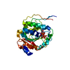
| ||||||||
|---|---|---|---|---|---|---|---|---|---|
| 1 | 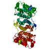
| ||||||||
| Unit cell |
|
-Components
| #1: Protein | Mass: 35938.012 Da / Num. of mol.: 1 Source method: isolated from a genetically manipulated source Source: (gene. exp.) Mycobacterium tuberculosis (bacteria) / Gene: MT1246, Rv1208 / Plasmid: pET30a / Production host: References: UniProt: O05309, UniProt: P9WMW9*PLUS, Transferases; Glycosyltransferases; Hexosyltransferases |
|---|---|
| #2: Chemical | ChemComp-MG /   Mass: 24.305 Da / Num. of mol.: 1 / Source method: obtained synthetically / Formula: Mg Mass: 24.305 Da / Num. of mol.: 1 / Source method: obtained synthetically / Formula: Mg |
| #3: Chemical | ChemComp-UDP /   Type: RNA linking / Mass: 404.161 Da / Num. of mol.: 1 / Source method: obtained synthetically / Formula: C9H14N2O12P2 / Comment: UDP*YM Type: RNA linking / Mass: 404.161 Da / Num. of mol.: 1 / Source method: obtained synthetically / Formula: C9H14N2O12P2 / Comment: UDP*YM |
| #4: Chemical | ChemComp-3PG /   Mass: 186.057 Da / Num. of mol.: 1 / Source method: obtained synthetically / Formula: C3H7O7P Mass: 186.057 Da / Num. of mol.: 1 / Source method: obtained synthetically / Formula: C3H7O7P |
| #5: Water | ChemComp-HOH /  Mass: 18.015 Da / Num. of mol.: 85 / Source method: isolated from a natural source / Formula: H2O Mass: 18.015 Da / Num. of mol.: 85 / Source method: isolated from a natural source / Formula: H2O |
-Experimental details
-Experiment
| Experiment | Method: X-RAY DIFFRACTION / Number of used crystals: 1 |
|---|
- Sample preparation
| Crystal | Density Matthews: 4.45 Å3/Da / Density % sol: 72.33 % |
|---|---|
| Crystal grow | Temperature: 293 K / Method: vapor diffusion, hanging drop / pH: 8 Details: 0.1M Tris pH 8.0, 0.5% polyethyleneglycol monomethyl ether 5000, 0.65M Na-K phosphate tetrahydrate, VAPOR DIFFUSION, HANGING DROP, temperature 293K |
-Data collection
| Diffraction | Mean temperature: 100 K |
|---|---|
| Diffraction source | Source: SYNCHROTRON / Site: ESRF  / Beamline: ID14-3 / Wavelength: 0.931 Å / Beamline: ID14-3 / Wavelength: 0.931 Å |
| Detector | Type: ADSC Q4R / Detector: CCD / Date: Jul 1, 2007 |
| Radiation | Monochromator: Diamond (111), Ge(220) / Protocol: SINGLE WAVELENGTH / Monochromatic (M) / Laue (L): M / Scattering type: x-ray |
| Radiation wavelength | Wavelength: 0.931 Å / Relative weight: 1 |
| Reflection | Resolution: 2.7→70.9 Å / Num. all: 17283 / Num. obs: 16855 / % possible obs: 97.6 % / Observed criterion σ(F): 0 / Observed criterion σ(I): 0 / Redundancy: 3.2 % / Biso Wilson estimate: 42.21 Å2 / Rmerge(I) obs: 0.082 / Rsym value: 0.069 / Net I/σ(I): 9.5 |
| Reflection shell | Resolution: 2.7→2.85 Å / Redundancy: 3.2 % / Rmerge(I) obs: 0.394 / Mean I/σ(I) obs: 2.6 / Num. unique all: 2481 / Rsym value: 0.33 / % possible all: 98.7 |
- Processing
| Software |
| |||||||||||||||||||||||||||||||||||||||||||||||||
|---|---|---|---|---|---|---|---|---|---|---|---|---|---|---|---|---|---|---|---|---|---|---|---|---|---|---|---|---|---|---|---|---|---|---|---|---|---|---|---|---|---|---|---|---|---|---|---|---|---|---|
| Refinement | Method to determine structure: MOLECULAR REPLACEMENT Starting model: Rubrobacter xylanophilus mannosyl-3-phosphoglycerate synthase (Sa-Moura et al., Acta Cryst. (2008). F64, 760-763) Resolution: 2.7→32.335 Å / Occupancy max: 1 / Occupancy min: 0.66 / SU ML: 0.34 / Cross valid method: THROUGHOUT / σ(F): 0 / σ(I): 0 / Phase error: 25.28 / Stereochemistry target values: ML
| |||||||||||||||||||||||||||||||||||||||||||||||||
| Solvent computation | Shrinkage radii: 0.9 Å / VDW probe radii: 1.11 Å / Solvent model: FLAT BULK SOLVENT MODEL / Bsol: 32.422 Å2 / ksol: 0.324 e/Å3 | |||||||||||||||||||||||||||||||||||||||||||||||||
| Displacement parameters | Biso max: 115.71 Å2 / Biso mean: 45.122 Å2 / Biso min: 23.94 Å2
| |||||||||||||||||||||||||||||||||||||||||||||||||
| Refinement step | Cycle: LAST / Resolution: 2.7→32.335 Å
| |||||||||||||||||||||||||||||||||||||||||||||||||
| Refine LS restraints |
| |||||||||||||||||||||||||||||||||||||||||||||||||
| LS refinement shell | Refine-ID: X-RAY DIFFRACTION / Total num. of bins used: 6
|
