 Movie
Movie Controller

[English] 日本語
 Yorodumi
- PDB-5juc: Crystal structure of glucosyl-3-phosphoglycerate synthase from My... -
+ Open data

- Basic information
| Entry | Database: PDB / ID: 5juc | ||||||
|---|---|---|---|---|---|---|---|
| Title | Crystal structure of glucosyl-3-phosphoglycerate synthase from Mycobacterium tuberculosis in complex with Mn2+, uridine-diphosphate (UDP) and glucosyl-3-phosphoglycerate (GPG) - GpgS*GPG*UDP*Mn2+_2 | ||||||
 Components Components | Glucosyl-3-phosphoglycerate synthase | ||||||
 Keywords Keywords | TRANSFERASE | ||||||
| Function / homology |  Function and homology information Function and homology informationglucosyl-3-phosphoglycerate synthase / UDP-alpha-D-glucose metabolic process / hexosyltransferase activity / magnesium ion binding / protein homodimerization activity Similarity search - Function | ||||||
| Biological species |   Mycobacterium tuberculosis (bacteria) Mycobacterium tuberculosis (bacteria) | ||||||
| Method |  X-RAY DIFFRACTION / SYNCHROTRON / MOLECULAR REPLACEMENT / Resolution: 2.8 Å X-RAY DIFFRACTION / SYNCHROTRON / MOLECULAR REPLACEMENT / Resolution: 2.8 Å | ||||||
 Authors Authors | Albesa-Jove, D. / Sancho-Vaello, E. / Rodrigo-Unzueta, A. / Comino, N. / Carreras-Gonzalez, A. / Arrasate, P. / Urresti, S. / Guerin, M.E. | ||||||
 Citation Citation | Journal: Structure / Year: 2017 Title: Structural Snapshots and Loop Dynamics along the Catalytic Cycle of Glycosyltransferase GpgS. Authors: Albesa-Jove, D. / Romero-Garcia, J. / Sancho-Vaello, E. / Contreras, F.X. / Rodrigo-Unzueta, A. / Comino, N. / Carreras-Gonzalez, A. / Arrasate, P. / Urresti, S. / Biarnes, X. / Planas, A. / Guerin, M.E. | ||||||
| History |
|
- Structure visualization
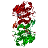
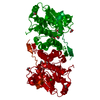
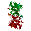
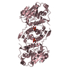
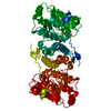
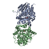
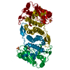
| Structure viewer | Molecule: MolmilJmol/JSmol |
|---|
- Downloads & links
-Download
| PDBx/mmCIF format | 5juc.cif.gz | 70.4 KB | Display | PDBx/mmCIF format |
|---|---|---|---|---|
| PDB format | pdb5juc.ent.gz | 49.8 KB | Display | PDB format |
| PDBx/mmJSON format | 5juc.json.gz | Tree view | PDBx/mmJSON format | |
| Others |  Other downloads Other downloads |
-Validation report
| Arichive directory | https://data.pdbj.org/pub/pdb/validation_reports/ju/5jucftp://data.pdbj.org/pub/pdb/validation_reports/ju/5juc | HTTPS FTP |
|---|
-Related structure data
| Related structure data | 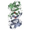 5jqxC 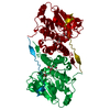 5jsxC 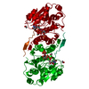 5jt0C 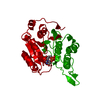 5judC 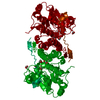 4y6nS C: citing same article ( S: Starting model for refinement |
|---|---|
| Similar structure data |


-Links
 PDBj
PDBj


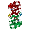
- Assembly
| Deposited unit | 
| ||||||||
|---|---|---|---|---|---|---|---|---|---|
| 1 | 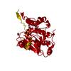
| ||||||||
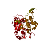
| Unit cell |
|
-Components
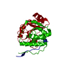
-Protein / Sugars , 2 types, 2 molecules A
| #1: Protein | Mass: 34683.555 Da / Num. of mol.: 1 Source method: isolated from a genetically manipulated source Source: (gene. exp.) Mycobacterium tuberculosis (strain ATCC 25618 / H37Rv) (bacteria)Gene: gpgS, Rv1208 / Production host: References: UniProt: P9WMW9, glucosyl-3-phosphoglycerate synthase |
|---|---|
| #5: Sugar | ChemComp-XDX / ( Type: D-saccharide / Mass: 348.198 Da / Num. of mol.: 1 / Source method: obtained synthetically / Formula: C9H17O12P Type: D-saccharide / Mass: 348.198 Da / Num. of mol.: 1 / Source method: obtained synthetically / Formula: C9H17O12P |
-Non-polymers , 4 types, 33 molecules 


| #2: Chemical | ChemComp-UDP /  Type: RNA linking / Mass: 404.161 Da / Num. of mol.: 1 / Source method: obtained synthetically / Formula: C9H14N2O12P2 / Comment: UDP*YM Type: RNA linking / Mass: 404.161 Da / Num. of mol.: 1 / Source method: obtained synthetically / Formula: C9H14N2O12P2 / Comment: UDP*YM | ||||
|---|---|---|---|---|---|
| #3: Chemical |  Mass: 62.068 Da / Num. of mol.: 2 / Source method: obtained synthetically / Formula: C2H6O2 Mass: 62.068 Da / Num. of mol.: 2 / Source method: obtained synthetically / Formula: C2H6O2#4: Chemical | ChemComp-MN / |  Mass: 54.938 Da / Num. of mol.: 1 / Source method: obtained synthetically / Formula: Mn Mass: 54.938 Da / Num. of mol.: 1 / Source method: obtained synthetically / Formula: Mn#6: Water | ChemComp-HOH / | Mass: 18.015 Da / Num. of mol.: 29 / Source method: isolated from a natural source / Formula: H2O |
-Experimental details
-Experiment
| Experiment | Method: X-RAY DIFFRACTION / Number of used crystals: 1 |
|---|
- Sample preparation
| Crystal | Density Matthews: 4.52 Å3/Da / Density % sol: 72.77 % |
|---|---|
| Crystal grow | Temperature: 291 K / Method: vapor diffusion, hanging drop / pH: 7.5 / Details: 14% PEG 8,000, 0.3-0.5 M Li sulfate |
-Data collection
| Diffraction | Mean temperature: 100 K |
|---|---|
| Diffraction source | Source: SYNCHROTRON / Site: Diamond  / Beamline: I03 / Wavelength: 0.97933 Å / Beamline: I03 / Wavelength: 0.97933 Å |
| Detector | Type: DECTRIS PILATUS3 6M / Detector: PIXEL / Date: Feb 27, 2015 |
| Radiation | Protocol: SINGLE WAVELENGTH / Monochromatic (M) / Laue (L): M / Scattering type: x-ray |
| Radiation wavelength | Wavelength: 0.97933 Å / Relative weight: 1 |
| Reflection | Resolution: 2.55→70.32 Å / Num. obs: 20094 / % possible obs: 99 % / Redundancy: 6.5 % / Biso Wilson estimate: 59.3 Å2 / CC1/2: 0.998 / Rmerge(I) obs: 0.1158 / Net I/σ(I): 7.94 |
| Reflection shell | Resolution: 2.55→2.641 Å / Redundancy: 6.5 % / Mean I/σ(I) obs: 1.15 / % possible all: 100 |
- Processing
| Software |
| |||||||||||||||||||||||||||||||||||||||||||||||||
|---|---|---|---|---|---|---|---|---|---|---|---|---|---|---|---|---|---|---|---|---|---|---|---|---|---|---|---|---|---|---|---|---|---|---|---|---|---|---|---|---|---|---|---|---|---|---|---|---|---|---|
| Refinement | Method to determine structure: MOLECULAR REPLACEMENT Starting model: 4Y6N Resolution: 2.8→70.31 Å / SU ML: 0.38 / Cross valid method: FREE R-VALUE / σ(F): 1.34 / Phase error: 24.51 / Stereochemistry target values: ML
| |||||||||||||||||||||||||||||||||||||||||||||||||
| Solvent computation | Shrinkage radii: 0.9 Å / VDW probe radii: 1.11 Å / Solvent model: FLAT BULK SOLVENT MODEL | |||||||||||||||||||||||||||||||||||||||||||||||||
| Refinement step | Cycle: LAST / Resolution: 2.8→70.31 Å
| |||||||||||||||||||||||||||||||||||||||||||||||||
| Refine LS restraints |
| |||||||||||||||||||||||||||||||||||||||||||||||||
| LS refinement shell |
|
